Talin-1 Antibody (YQ-16)santacruz SC-81805

Talin-1 Antibody (YQ-16)

  • 产品型号:  santacruz SC-81805
  • 简单描述
  • mouse monoclonal IgG3 provided at 100 µg/mlrecommended for detection of Talin-1 of mouse, rat and human origin by WB, IP, IF, IHC(P) and ELISA
详细介绍

SANTA CRUZ BIOTECHNOLOGY, INC.
Talin-1 (YQ-16): sc-81805
Santa Cruz Biotechnology, Inc. 1.800.457.3801 831.457.3800 831.457.3801 Europe +00800 4573 8000 49 6221 4503 0 www.scbt.com
BACKGROUND
Focal adhesions were identified as areas within the plasma membrane of
tissue culture cells that adhere tightly to the underlying substrate. In vivo,
these regions are involved in the adhesion of cells to the extracellular matrix.
Paxillin and vinculin are cytoskeletal, focal adhesion proteins that are components
of a protein complex that links the Actin network to the plasma
membrane. Vinculin binding sites have been identified on other cytoskeletal
proteins, including Talin-1 and α-actinin. In addition, vinculin, Talin-1, Talin-2
and α-actinin each contain Actin binding sites. Expression of vinculin, Talin-1
and Talin-2 have been shown to be affected by the level of Actin expression.
α-actinin has been shown to link Actin to integrins in the plasma membrane
through interactions with the vinculin and Talin complex or by a direct interaction
with integrin. Talin-2 is similar to Talin-1 but shows distinct patterns of
expression and cannot compensate for the loss of Talin-1.
REFERENCES
1. Burridge, K., et al. 1988. Focal adhesions: transmembrane junctions
between the extracellular matrix and the cytoskeleton. Annu. Rev. Cell
Biol. 4: 487-525.
2. Gilmore, A.P., et al. 1992. Further characterization of the Talin-binding site
in the cytoskeletal protein vinculin. J. Cell Sci. 103: 719-731.
3.Wood, C.K., et al. 1994. Characterisation of the paxillin-binding site and
the C-terminal focal adhesion targeting sequence in vinculin. J. Cell Sci.
107: 709-717.
4. Gluck, U., et al. 1994. Modulation of α-actinin levels affects cell motility
and confers tumorigenicity on 3T3 cells. J. Cell Sci. 107: 1773-1782.
5. Schevzov, G., et al. 1995. Impact of Actin gene expression on vinculin,
Talin, cell spreading, and motility. DNA Cell Biol. 14: 689-700.
6. Hemmings, L., et al. 1996. Talin contains three Actin-binding sites each of
which is adjacent to a vinculin-binding site. J. Cell Sci. 109: 2715-2726.
7. Gilmore, A.P., et al. 1996. Regulation of vinculin binding to Talin and Actin
by phosphatidyl-inositol-4-5-bisphosphate. Nature 381: 531-535.
CHROMOSOMAL LOCATION
Genetic locus: TLN1 (human) mapping to 9p13.3; Tln1 (mouse) mapping to
4 B1.
SOURCE
Talin-1 (YQ-16) is a mouse monoclonal antibody raised against recombinant
Talin-1 of human origin.
PRODUCT
Each vial contains 100 μg IgG3 in 1.0 ml PBS with < 0.1% sodium azide and
0.1% gelatin.
STORAGE
Store at 4° C, **DO NOT FREEZE**. Stable for one year from the date of
shipment. Non-hazardous. No MSDS required.
APPLICATIONS
Talin-1 (YQ-16) is recommended for detection of Talin-1 of mouse, rat and
human origin by Western Blotting (starting dilution 1:200, dilution range
1:100-1:1000), immunoprecipitation [1-2 μg per 100-500 μg of total protein
(1 ml of cell lysate)], immunofluorescence (starting dilution 1:50, dilution
range 1:50-1:500), immunohistochemistry (including paraffin-embedded
sections) (starting dilution 1:50, dilution range 1:50-1:500) and solid phase
ELISA (starting dilution 1:30, dilution range 1:30-1:3000).
Suitable for use as control antibody for Talin-1 siRNA (h): sc-36610, Talin-1
siRNA (m): sc-36611, Talin-1 shRNA Plasmid (h): sc-36610-SH, Talin-1 shRNA
Plasmid (m): sc-36611-SH, Talin-1 shRNA (h) Lentiviral Particles: sc-36610-V
and Talin-1 shRNA (m) Lentiviral Particles: sc-36611-V.
Molecular Weight of Talin-1: 230 kDa.
Positive Controls: RAW 264.7 whole cell lysate: sc-2211.
RECOMMENDED SECONDARY REAGENTS
To ensure optimal results, the following support (secondary) reagents are
recommended: 1) Western Blotting: use goat anti-mouse IgG-HRP: sc-2005
(dilution range: 1:2000-1:32,000) or Cruz Marker™ compatible goat antimouse
IgG-HRP: sc-2031 (dilution range: 1:2000-1:5000), Cruz Marker™
Molecular Weight Standards: sc-2035, TBS Blotto A Blocking Reagent:
sc-2333 and Western Blotting Luminol Reagent: sc-2048. 2) Immunoprecipitation:
use Protein A/G PLUS-Agarose: sc-2003 (0.5 ml agarose/2.0 ml).
3) Immunofluorescence: use goat anti-mouse IgG-FITC: sc-2010 (dilution
range: 1:100-1:400) or goat anti-mouse IgG-TR: sc-2781 (dilution range:
1:100-1:400) with UltraCruz™ Mounting Medium: sc-24941. 4) Immunohistochemistry:
use ImmunoCruz™: sc-2050 or ABC: sc-2017 mouse IgG
Staining Systems.
DATA
SELECT PRODUCT CITATIONS
1. Ah Kioon, M.D., et al. 2010. Adrenomedullin increases fibroblast-like synoviocyte
adhesion to extracellular matrix proteins by upregulating integrin
activation. Arthritis Res. Ther. 12: R190.
RESEARCH USE
For research use only, not for use in diagnostic procedures.


santacruz GSK-3 Inhibitor XSC-221689

santacruz GSK-3 Inhibitor X

  • 产品型号:  SC-221689
  • 简单描述
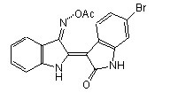
  • santacruz GSK-3 Inhibitor X分子量: 398.2分子式: C18H12BrN3O3可溶性: Soluble in DMSO
详细介绍

santacruz GSK-3 Inhibitor X
详细产品信息:

GSK-3 Inhibitor X (CAS 740841-15-0)

5 5santacruz GSK-3 Inhibitor XSC-221689santacruz GSK-3 Inhibitor XSC-221689santacruz GSK-3 Inhibitor XSC-221689santacruz GSK-3 Inhibitor XSC-221689santacruz GSK-3 Inhibitor XSC-221689 (Based on popularity) 1

santacruz GSK-3 Inhibitor XSC-221689

Synonym: BIO-Acetoxime; 6-Bromoindirubin-3′-acetoxime, BIA
Application: A selective GSK-3α/β inhibitor
CAS Number: 740841-15-0
Purity: ≥95%
Molecular Weight: 398.21
Molecular Formula: C18H12BrN3O3
Refer to Certificate of Analysis for lot specific data (including water content).
Set Currency US DollarsEuroSwiss FrancsBritish PoundsAustralia DollarsCanadian DollarsRMB Add Selected


 Ordering Information

Product Name Catalog # Unit Price Qty Add Favorites
GSK-3 Inhibitor X sc-221689 1 mg $115   Add  

Description

GSK-3 Inhibitor X is an acetoxime analog of BIO which acts as a selective GSK-3α/β inhibitor (IC50 values are 0.01, 2.4, 4.3 and 63 μM for GSK-3α/β, CDK5/p25, CDK2/cyclin A and CDK1/cyclin B respectively). GSK-3 Inhibitor X induces Wnt signaling and inhibits CD8+ T cell effector differentiation.

Technical Information

Physical State: Solid
Solubility: Soluble in DMSO (10 mg/ml).
Storage: Store at 4° C
Melting Point: 236.83 °C (Predicted)
Boiling Point: 551.87 °C (Predicted)
Density: 1.69 g/cm3 (Predicted)
Refractive Index: n20D 1.74 (Predicted)
IC50: GSK-3α/β: IC50 = 10 nM; Cdk5/p25: IC50 = 2.4 µM; Cdk2/A: IC50 = 4.3 µM; Cdk1/B: IC50 = 63 µM; Cdk4/D1: IC50 = ≥10 µM; aryl hydrocarbon receptor: EC5050 = ≥0.1 µM (yeast); aryl hydrocarbon receptor: EC5050 = 0.16 µM (mammalian)
pK Values: pKa: 9.23 (Predicted)

Safety and Reference Information

PubChem CID: 6538818
MDL Number: MFCD09037533
SMILES: CC(=O)ONC1=C2C=CC=CC2=NC1=C3C4=C(C=C(C=C4)Br)NC3=O

For Research Use Only. Not Intended for Diagnostic or Therapeutic Use.


详细产品信息也可和选购


Santacruz抗体代理SOCS-1 (C-20)-R AntibodySC-7005-R

Santacruz抗体代理SOCS-1 (C-20)-R Antibody

  • 产品型号:  SC-7005-R
  • 简单描述
  • Santacruz抗体代理SOCS-1 (C-20)-R Antibody rabbit polyclonal IgG, 200 ?g/mlepitope mapping at the C-terminus of SOCS-1 of human originrecommended for detection of SOCS-1 of mouse, rat and human origi
详细介绍


还有同类其他抗原和反应种属的其他产品,欢饮!


SOCS-2 Antibody (H-74)Santacruz抗体供应santacruz代理 SC-9022

SOCS-2 Antibody (H-74)Santacruz抗体供应

  • 产品型号:  santacruz代理 SC-9022
  • 简单描述
  • •rabbit polyclonal IgG, 200 µg/mlWB IP IF IHC(P) siRNA •epitope corresponding to amino acids 89-162 mapping within an internal region of SOCS-2 of human origin
详细介绍

此类产品还有其他抗体来源和种属反应详细产品信息请,谢谢!


santacruz G蛋白PLUS-琼脂糖Protein G PLUS-AgaroseSC-2002

santacruz G蛋白PLUS-琼脂糖Protein G PLUS-Agarose

  • 产品型号:  SC-2002
  • 简单描述
  • Protein G PLUS-Agarose is suitable for immunoprecipitation of mouse IgG1,IgG2a, IgG2b and IgG3, rat IgG1, IgG2a, santacruz G蛋白PLUS-琼脂糖Protein G PLUS-AgaroseIgG2b and IgG2c, rabbit and goat polycl
详细介绍

santacruz  G蛋白PLUS-琼脂糖Protein G PLUS-Agarose

SANTA CRUZ BIOTECHNOLOGY, INC.
Protein G PLUS-Agarose
Immunoprecipitation Reagent: sc-2002
Santa Cruz Biotechnology, Inc. 1.800.457.3801 831.457.3800 831.457.3801 Europe +00800 4573 8000 49 6221 4503 0 www.scbt.com
PRODUCT
Protein G PLUS is provided as an agarose conjugate for use in immunoprecipitation
only. The product is provided as 0.5 ml agarose in 2.0 ml PBS buffer
with 0.02% azide. Protein G PLUS-Agarose is pre-blocked with BSA to reduce
non-specific immunoglobulin binding. Sufficient product is provided for 100
immunoprecipitation reactions, to be used at 20 μl resuspended volume per
reaction.
SPECIFICITY
Protein G PLUS-Agarose is suitable for immunoprecipitation of mouse IgG1,
IgG2a, IgG2b and IgG3, rat IgG1, IgG2a, IgG2b and IgG2c, rabbit and goat polyclonal
Abs, and human IgG1, IgG2, IgG3 and IgG4.
PROCEDURE
• Incubate cultured cells (80-90% confluent monolayer in 100 mm cell culture
plate, or approximay 2-5 x 10⁷ suspension cells in flask) in methioninefree
medium containing 5% dialyzed fetal calf serum for 1 hour at 37° C.
The same procedure can be used for cells labeled with other radioactive
amino acids (e.g., 14C or 3H) or with γ32P-orthophosphate. Cell labeling must
be carried out in medium lacking the relevant amino acid or in phosphatefree
medium.
• Remove medium and replace with 3 ml methionine-free medium containing
5% dialyzed fetal calf serum and 100 μCi/ml 35S-methionine. Incubate 1
hour at 37° C. For some proteins a longer labeling period (up to 18 hours)
is preferable.
• Carefully remove radioactive medium with Pasteur pipette and wash cell
monolayer with PBS.
• Add 3 ml ice cold RIPA buffer to cell monolayer and incubate at 4° C for
10 minutes. For suspension cells, add the RIPA buffer to washed cell pellet
in a 15 ml conical centrifuge tube.
• Disrupt cells by repeated aspiration through a 21 gauge needle and transfer
to a 15 ml conical centrifuge tube.
• Wash cell culture plate with additional 1.0 ml ice cold RIPA buffer and
combine with original extract.
• Pellet cellular debris by centrifugation at 10,000 xg for 10 minutes at 4° C.
Transfer supernatant to a fresh 15 ml conical centrifuge tube on ice.
Preclear lysate (optional step) by adding 1.0 μg of the appropriate control
IgG (normal mouse, rat, rabbit or goat IgG, corresponding to the host
species of the primary antibody), together with 20 μl of resuspended
volume of Protein G PLUS-Agarose. Incubate at 4° C for 30 minutes.
• Pellet beads by centrifugation at 2,500 rpm (approximay 1,000 xg) for
5 minutes at 4° C. Transfer supernatant (cell lysate) to a fresh 15 ml conical
centrifuge tube on ice.
• Transfer 1 ml of the above cell lysate, or approximay 100-500 μg total
cellular protein, to a 1.5 ml microcentrifuge tube. Add 1-10 μl (i.e., 0.2-2 μg)
primary antibody (optimal antibody concentration should be determined by
titration) and incubate for 1 hour at 4° C.
• Add 20 μl of resuspended volume of Protein G PLUS-Agarose. Cap tubes
and incubate at 4° C on a rocker platform or rotating device for 1 hour to
overnight.
• Collect immunoprecipitates by centrifugation at 2,500 rpm (approximay
1,000 xg) for 5 minutes at 4° C. Carefully aspirate and discard radioactive
supernatant.
• Wash pellet 4 times with 1.0 ml RIPA buffer (more stringent) or PBS (less
stringent), each time repeating centrifugation step above.
• After final wash, aspirate and discard supernatant and resuspend pellet in
40 μl of 1x electrophoresis sample buffer.
• Boil samples for 2–3 minutes and analyze 20 μl aliquots by SDS-PAGE and
autoradiography. Unused samples may be stored at -20° C.
• Optional: After boiling, samples may be centrifuged to pellet the agarose
beads followed by SDS-PAGE analysis of the supernatant.
SELECT PRODUCT CITATIONS
1. Medema, R., et al. 1998. p21waf1 can block cells at two points in the cell
cycle, but does not interfere with processive DNA-replication or stressactivated
kinases. Oncogene 16: 431-441.
2. Marzo, I., et al. 1998. Bax and adenine nucleotide translocator cooperate
in the mitochondrial control of apoptosis. Science 281: 2027-2031.
3. Song, Y., et al. 2011. Ligand-dependent corepressor acts as a novel corepressor
of thyroid hormone receptor and represses hepatic lipogenesis in
mice. J. Hepatol. 56: 248-254.
4. Beard, R.S., et al. 2012. Metabotropic glutamate receptor 5 mediates
phosphorylation of vascular endothelial cadherin and nuclear localization
of β-catenin in response to homocysteine. Vascul. Pharmacol. 56: 159-167.
STORAGE
Store at 4° C, do not freeze; stable for one year from the date of shipment.
RESEARCH USE
For research use only, not for use in diagnostic procedures.
Immunoprecipitation agarose conjugates are pre-blocked with BSA to reduce non-specific immunoglobulin
binding and are provided at a concentration (0.5 ml agarose/2.0 ml) suitable for use at 20 μl per
immunoprecipitation reaction. Number of reactions: 100.


santacruz PCGF6 (E-15) 抗体*SC-160649

santacruz PCGF6 (E-15) 抗体*

  • 产品型号:  SC-160649
  • 简单描述
  • santacruz PCGF6 (E-15) 抗体*Epitope: Internal (h) Applications: WB, IF, ELISA Species: m, r, hIsotype: goat IgG
详细介绍

santacruz PCGF6 (E-15) 抗体*

BACKGROUND
Polycomb group (PcG) proteins form multiprotein complexes that regulate
expression patterns of developmental and cell proliferation genes. Several
members of the PcG contain ring finger domains and are identified as a subclass
of RING finger proteins. The RING-type zinc finger motif is present in a
number of viral and eukaryotic proteins and is made of a conserved cysteinerich
domain that is able to bind two zinc atoms. Proteins that contain the
RING-type zinc finger conserved domain are generally involved in the ubiquitination
pathway of protein degradation. PCGF6 (polycomb group ring finger
6), also known as MBLR or RNF134, is a 350 amino acid nuclear protein
that is ubiquitously expressed and contains one RING-type zinc finger. PCGF6
acts as a transcriptional repressor and regulates the level of histone
H3K4Me3 by activating SmcY histone demethylase. PCGF6 exists as two
alternatively spliced isoforms.
REFERENCES
1. Kanno, M., Hasegawa, M., Ishida, A., Isono, K. and Taniguchi, M. 1995.
Mel-18, a Polycomb group-related mammalian gene, encodes a transcriptional
negative regulator with tumor suppressive activity. EMBO J. 14:
5672-5678.
2. Satijn, D.P., Gunster, M.J., van der Vlag, J., Hamer, K.M., Schul, W.,
Alkema, M.J., Saurin, A.J., Freemont, P.S., van Driel, R. and Otte, A.P.
1997. RING1 is associated with the polycomb group protein complex and
acts as a transcriptional repressor. Mol. Cell. Biol. 17: 4105-4113.
3. Joazeiro, C.A. and Weissman, A.M. 2000. RING finger proteins: mediators
of ubiquitin ligase activity. Cell 102: 549-552.
4. Akasaka, T., Takahashi, N., Suzuki, M., Koseki, H., Bodmer, R. and Koga, H.
2002. MBLR, a new RING finger protein resembling mammalian Polycomb
gene products, is regulated by cell cycle-dependent phosphorylation. Genes
Cells 7: 835-850.
5. Tuckfield, A., Clouston, D.R., Wilanowski, T.M., Zhao, L.L., Cunningham,
J.M. and Jane, S.M. 2002. Binding of the RING polycomb proteins to specific
target genes in complex with the grainyhead-like family of developmental
transcription factors. Mol. Cell. Biol. 22: 1936-1946.
6. Online Mendelian Inheritance in Man, OMIM™. 2003. Johns Hopkins
University, Baltimore, MD. MIM Number: 607816. World Wide Web URL:
http://www.ncbi.nlm.nih.gov/omim/
7. Moore, R. and Boyd, L. 2004. Analysis of RING finger genes required for
embryogenesis in C. elegans. Genesis 38: 1-12.
8. Yang, Y., Lorick, K.L., Jensen, J.P. and Weissman, A.M. 2005. Expression
and evaluation of RING finger proteins. Meth. Enzymol. 398: 103-112.
9. Lee, M.G., Norman, J., Shilatifard, A. and Shiekhattar, R. 2007. Physical and
functional association of a trimethyl H3K4 demethylase and Ring6a/MBLR,
a polycomb-like protein. Cell 128: 877-887.
STORAGE
Store at 4° C, **DO NOT FREEZE**. Stable for one year from the date of
shipment. Non-hazardous. No MSDS required.
CHROMOSOMAL LOCATION
Genetic locus: PCGF6 (human) mapping to 10q24.33; Pcgf6 (mouse) mapping
to 19 C3.
SOURCE
PCGF6 (E-15) is an affinity purified goat polyclonal antibody raised against a
peptide mapping within an internal region of PCGF6 of human origin.
PRODUCT
Each vial contains 200 μg IgG in 1.0 ml of PBS with < 0.1% sodium azide
and 0.1% gelatin.
Blocking peptide available for competition studies, sc-160649 P, (100 μg
peptide in 0.5 ml PBS containing < 0.1% sodium azide and 0.2% BSA).
APPLICATIONS
PCGF6 (E-15) is recommended for detection of PCGF6 of mouse, rat and
human origin by Western Blotting (starting dilution 1:200, dilution range
1:100-1:1000), immunofluorescence (starting dilution 1:50, dilution range
1:50-1:500) and solid phase ELISA (starting dilution 1:30, dilution range
1:30-1:3000); non cross-reactive with PCGF1, PCGF3 or PCGF5.
Suitable for use as control antibody for PCGF6 siRNA (h): sc-90663, PCGF6
siRNA (m): sc-152109, PCGF6 shRNA Plasmid (h): sc-90663-SH, PCGF6
shRNA Plasmid (m): sc-152109-SH, PCGF6 shRNA (h) Lentiviral Particles:
sc-90663-V and PCGF6 shRNA (m) Lentiviral Particles: sc-152109-V.
Molecular Weight of PCGF6: 60 kDa.
RECOMMENDED SECONDARY REAGENTS
To ensure optimal results, the following support (secondary) reagents are
recommended: 1) Western Blotting: use donkey anti-goat IgG-HRP: sc-2020
(dilution range: 1:2000-1:100,000) or Cruz Marker™ compatible donkey
anti-goat IgG-HRP: sc-2033 (dilution range: 1:2000-1:5000), Cruz Marker™
Molecular Weight Standards: sc-2035, TBS Blotto A Blocking Reagent:
sc-2333 and Western Blotting Luminol Reagent: sc-2048. 2) Immunofluorescence:
use donkey anti-goat IgG-FITC: sc-2024 (dilution range: 1:100-
1:400) or donkey anti-goat IgG-TR: sc-2783 (dilution range: 1:100-1:400)
with UltraCruz™ Mounting Medium: sc-24941.
RESEARCH USE
For research use only, not for use in diagnostic procedures.
PROTOCOLS