Anti-Mouse F4/80 Antigen PE 抗小鼠F4/80抗原PE抗体eBiosience12-4801-82

Anti-Mouse F4/80 Antigen PE 抗小鼠F4/80抗原PE抗体

  • 产品型号:  eBiosience12-4801-82
  • 简单描述
  • HostRatIsotypeIgG2a, kappaReactivityMouseConjugatePELaserBlue Laser, Green Laser, Yellow-Green LaserEmit575 nmExcite488 – 561 nmReported ApplicationsFlow Cytometric Analysis
详细介绍

Also known as: Pan Macrophage Marker

Clone: BM8

Description: The BM8 monoclonal antibody reacts with mouse F4/80 antigen, an approximay 125 kDa transmembrane protein. The F4/80 antigen is expressed by a majority of mature macrophages and is the best marker for this population of cells. However, other cell types such as Langerhans cells and liver Kupffer cells have been reported to express this antigen. Expression of F4/80 commences during early myeloid development and is upregulated on all BM cells stimulated in vitro with M-CSF. It has been shown that some cytokines downregulate the expression of F4/80 resulting in lack of F4/80 antigen on a subpopulation of macrophages, especially in the lymphoid microenvironment in vivo

Host Rat
Isotype IgG2a, kappa
Reactivity Mouse
Conjugate PE
Laser Blue Laser, Green Laser, Yellow-Green Laser
Emit 575 nm
Excite 488 – 561 nm
Reported Applications Flow Cytometric Analysis

Anti-pan Cytokeratin antibody PCK-26 鼠单克隆[PCK-26abcam ab6401

Anti-pan Cytokeratin antibody PCK-26 鼠单克隆[PCK-26

  • 产品型号:  abcam ab6401
  • 简单描述
  • Anti-pan Cytokeratin antibody [PCK-26] 鼠单克隆[PCK-26DescriptionMouse monoclonal [PCK-26] to pan CytokeratinTested applicationsWB, IHC-FoFr, IHC-P, IHC-Fr, IF, Dot Blot, Flow Cyt, ICC/IFCro
详细介绍

Anti-pan Cytokeratin antibody [PCK-26] 鼠单克隆[PCK-26 

Form

Liquid

Storage instructions

Store at +4°C short term (1-2 weeks). Aliquot and store at -20°C or -80°C. Avoid repeated freeze / thaw cycles.

Storage buffer

Preservative: 15mM Sodium Azide
Constituents: Ascites

Concentration


Anti-pan Cytokeratin antibody [PCK-26] 鼠单克隆[PCK-26 


Purity

Ascites

Clonality

Monoclonal

Clone number

PCK-26

Myeloma

unknown

Isotype

IgG1

Light chain type

unknown

Research areas

Signal Transduction >> Cytoskeleton / ECM >> Cytoskeleton >> Intermediate Filaments >> Class I >> Cytokeratins
 

  • Datasheet PDF
  • SDS
  • Immunohistochemistry (Formalin/PFA-fixed paraffin-embedded sections) – pan Cytokeratin antibody [PCK-26] (ab6401)Anti-pan Cytokeratin antibody PCK-26 鼠单克隆[PCK-26abcam ab6401(enlarge)
  • Immunohistochemistry (Formalin/PFA-fixed paraffin-embedded sections) – pan Cytokeratin antibody [PCK-26] (ab6401)Anti-pan Cytokeratin antibody PCK-26 鼠单克隆[PCK-26abcam ab6401(enlarge)
  • Western blot – pan Cytokeratin antibody [PCK-26] (ab6401)Anti-pan Cytokeratin antibody PCK-26 鼠单克隆[PCK-26abcam ab6401(enlarge)
Applications

Show applications key

Our Abpromise guarantee covers the use of ab6401 in the following tested applications.

The application notes include recommended starting dilutions; optimal dilutions/concentrations should be determined by the end user.

  • ShowHide4 Images

    WB

    Four stars(5 Abreviews)WB: Use at an assay depe…Read more →

    WB: Use at an assay dependent dilution.

  • ShowHide

    IHC-FoFr

    IHC-FoFr: Use at an assa…Read more →

    IHC-FoFr: Use at an assay dependent dilution.

  • ShowHide3 Images

    IHC-P

    Five stars(3 Abreviews)IHC-P: Use at an assay d…Read more →

    IHC-P: Use at an assay dependent dilution.

  • ShowHide2 Images

    IHC-Fr

    Four stars(3 Abreviews)IHC-Fr: Use at an assay …Read more →

    IHC-Fr: Use at an assay dependent dilution.

  • IF

    IF: 1/300.

    IF: 1/300.

  • ShowHide

    Dot Blot

    Dot: Use at an assay dep…Read more →

    Dot: Use at an assay dependent dilution.

  • ShowHide

    Flow Cyt

    See more…Read more →

    Flow Cyt: Use at an assay dependent dilution. PubMed: 19855980Use 10µl for 5 x 10⁷ cells.

  • 2 Images

    ICC/IF

    Five stars(2 Abreviews)ICC/IF: 1/300.

    ICC/IF: 1/300.

Target

Relevance

Cytokeratins, a group comprising at least 29 different proteins, are characteristic of epithelial and trichocytic cells. Cytokeratins 1, 4, 5, 6, and 8 are members of the type II neutral to basic subfamily. Monoclonal anti cytokeratins are specific markers of epithelial cell differentiation and have been widely used as tools in tumor identification and classification. Monoclonal Anti Pan Cytokeratin (mixture) is a broadly reactive reagent, which recognizes epitopes present in most human epithelial tissues. It facilitates typing of normal, metaplastic and neoplastic cells. Synergy between the various components results in staining amplification. This enables identification of cells, which would otherwise be stained only marginally. The mixture may aid in the discrimination of carcinomas and nonepithelial tumors such as sarcomas, lymphomas and neural tumors. It is also useful in detecting micrometastases in lymph nodes, bone marrow and other tissues and for determining the origin of poorly differentiated tumors. There are two types of cytokeratins the acidic type I cytokeratins and the basic or neutral type II cytokeratins. Cytokeratins are usually found in pairs comprising a type I cytokeratin and a type II cytokeratin. Usually the type II cytokeratins are 8kD larger than their type I counterparts.

Cellular localization

Cytoplasmic

Anti-pan Cytokeratin antibody [PCK-26] images:

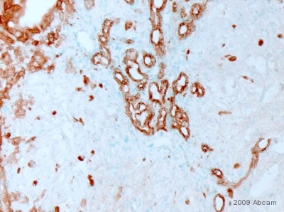
Immunohistochemistry (Formalin/PFA-fixed paraffin-embedded sections) – pan Cytokeratin antibody [PCK-26] (ab6401)
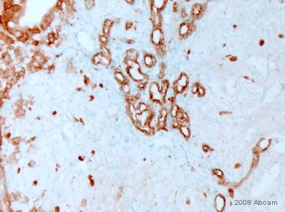
Anti-pan Cytokeratin antibody PCK-26 鼠单克隆[PCK-26abcam ab6401


ab6401 staining pan Cytokeratin in Mouse liver tumor tissue sections by IHC-P (Paraformaldehyde fixed paraffin embedded sections). Tissue was fixed with paraformaldehyde and blocked with maleate buffer blocking solution for 30 minutes and 22°C. Antigen retrieval was by heat mediation in citrate buffer. Samples were incubated with primary antibody (1/500) in maleate buffer blocking solution for 16 hours at 22°C. An undiluted biotin-conjugated Donkey polyclonal to mouse IgG was used as secondary antibody.

This image is courtesy of an Abreview submitted by Dr Asha Seth


See Abreview

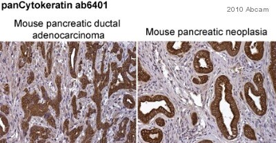
Immunohistochemistry (Formalin/PFA-fixed paraffin-embedded sections) – pan Cytokeratin antibody [PCK-26] (ab6401)
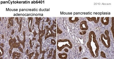
Anti-pan Cytokeratin antibody PCK-26 鼠单克隆[PCK-26abcam ab6401


ab6401 staining pan Cytokeratin in mouse pancreatic ductal adenocarcinoma (left-hand panel) and mouse pancreatic neoplasia (right-hand panel) sections by immunohistochemistry (IHC-P – paraformaldehyde-fixed, paraffin-embedded sections). Tissue samples were fixed with paraformaldehyde and blocked with 10% serum for 1 hour at room temperature; heat mediated antigen retrieval was performed. The sample was incubated with primary antibody (1/250) at 4°C for 8 hours. A Biotin-conjugated Goat polyclonal (1/1000) was used as the secondary antibody.

This image is courtesy of an anonymous Abreview


See Abreview

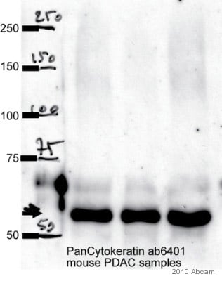
Western blot – pan Cytokeratin antibody [PCK-26] (ab6401)
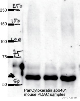
Anti-pan Cytokeratin antibody PCK-26 鼠单克隆[PCK-26abcam ab6401


Observed band size : 55 kDa (why is the actual band size different from the predicted?)

All lanes: pan Cytokeratin antibody (ab6401) at 1/1000 dilution + whole cell lysate of Primary mouse pancreatic cancer cell lines (35 µg)

Secondary: An HRP-conjugated Sheep anti-mouse IgG polyclonal (1/5000) developed using the ECL technique

Performed under non-reducing conditions.

Blocking Step: 10% Milk for 1 hour at room temperature

This image is courtesy of an Abreview submitted by Pawel Mazur


See Abreview

Immunohistochemistry (Frozen sections) – pan Cytokeratin antibody [PCK-26] (ab6401)
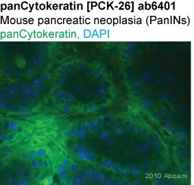
Anti-pan Cytokeratin antibody PCK-26 鼠单克隆[PCK-26abcam ab6401


ab6401 staining pan Cytokeratin in Mouse pancreatic neoplasia tissue sections by Immunohistochemistry (IHC-Fr – frozen sections). Tissue was fixed with paraformaldehyde and blocked with 1% BSA for 1 hour at room temperature. Samples were incubated with primary antibody (1/250 in PBS) for 8 hours at 4°C. An Alexa Fluor®488-conjugated goat anti-mouse IgG polyclonal (1/1000) was used as the secondary antibody.

This image is courtesy of an anonymous Abreview


See Abreview

Immunocytochemistry/ Immunofluorescence – pan Cytokeratin antibody [PCK-26] (ab6401)
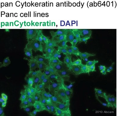
Anti-pan Cytokeratin antibody PCK-26 鼠单克隆[PCK-26abcam ab6401


ab6401 staining pan Cytokeratin in Human Panc1 cells by ICC/IF (Immunocytochemistry/immunofluorescence). Cells were fixed with acetone/methanol (1:1) and blocked with 1% BSA for 1 hour at room temperature. Samples were incubated with primary antibody (1/300 in PBS) for 2 hours. An Alexa Fluor®488-conjugated DGoat anti-mouse IgG polyclonal (1/1000) was used as the secondary antibody. Nuclei were counterstained with DAPI.

This image is courtesy of an anonymous Abreview


See Abreview

References for Anti-pan Cytokeratin antibody [PCK-26] (ab6401)


Anti-pan Cytokeratin antibody [PCK-26] 鼠单克隆[PCK-26 详细产品信息可和选购


Anti-CD11b antibody EP1345Y CD11兔单克隆抗体abcam ab52478

Anti-CD11b antibody EP1345Y CD11兔单克隆抗体

  • 产品型号:  abcam ab52478
  • 简单描述
  • Anti-CD11b antibody [EP1345Y] CD11兔单克隆抗体DescriptionRabbit monoclonal Tested applicationsWB, IP, ICC, Flow Cyt, IHC-PCross reactivityReacts withMouse, Human
详细介绍

Anti-CD11b antibody [EP1345Y] CD11兔单克隆抗体

Form

Liquid

Storage instructions

Shipped at 4°C. Upon delivery aliquot and store at -20°C. Avoid freeze / thaw cycles.

Storage buffer

PBS 49%,Sodium azide 0.01%,Glycerol 50%,BSA 0.05%

Purity

Tissue culture supernatant

Clonality

Monoclonal

Clone number

EP1345Y

Isotype

IgG

Research areas

Stem Cells >> Hematopoietic Progenitors >> Myeloid >> Neutrophil Lineage
Stem Cells >> Hematopoietic Progenitors >> Myeloid >> Monocytic Lineage
Stem Cells >> Hematopoietic Progenitors >> Lymphoid >> NK Cell Lineage
Stem Cells >> Hematopoietic Progenitors >> Lymphoid >> T Lymphocytic Lineage
Stem Cells >> Hematopoietic Progenitors >> Lymphoid >> B Lymphocytic Lineage
Stem Cells >> Hematopoietic Progenitors >> Hematopoietic Stem Cells >> Murine Lineage Negative
Stem Cells >> Mesenchymal Stem Cells >> Negative Markers
Neuroscience >> Cell Type Marker >> Glia marker >> Microglia marker
Tags & Cell Markers >> Cell Type Markers >> Neuroscience Markers >> Glial
Signal Transduction >> Cytoskeleton / ECM >> Cell Adhesion >> Integrins >> Alpha
 

  • Datasheet PDF
  • SDS
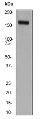
  • Western blot – CD11b antibody [EP1345Y] (ab52478)Anti-CD11b antibody EP1345Y CD11兔单克隆抗体abcam ab52478(enlarge)

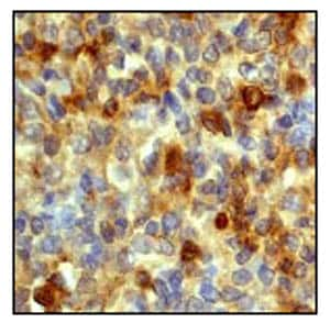
  • Immunohistochemistry (Paraffin-embedded sections) – CD11b antibody [EP1345Y] (ab52478)Anti-CD11b antibody EP1345Y CD11兔单克隆抗体abcam ab52478(enlarge)

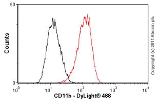
  • Flow Cytometry-CD11b antibody [EP1345Y](ab52478)Anti-CD11b antibody EP1345Y CD11兔单克隆抗体abcam ab52478(enlarge)


Anti-CD11b antibody [EP1345Y] CD11兔单克隆抗体

Applications

Show applications key

Our Abpromise guarantee covers the use of ab52478 in the following tested applications.

The application notes include recommended starting dilutions; optimal dilutions/concentrations should be determined by the end user.

  • ShowHide1 Image

    WB

    WB: 1/20000 – 1/50000. P…Read more →

    WB: 1/20000 – 1/50000. Predicted molecular weight: 128 kDa.

  • IP

    IP: 1/80.

    IP: 1/80.

  • ICC

    ICC: 1/100 – 1/250.

    ICC: 1/100 – 1/250.

  • 1 Image

    Flow Cyt

    Flow Cyt: 1/20 – 1/50.

    Flow Cyt: 1/20 – 1/50.

  • ShowHide1 Image

    IHC-P

Anti-CD11b antibody [EP1345Y] CD11兔单克隆抗体 详细产品信息可和选购

Anti-HE4 antibodyab24480

Anti-HE4 antibody

  • 产品型号:  ab24480
  • 简单描述
  • Anti-HE4 antibodyDescriptionRabbit polyclonal to HE4Tested applicationsWB, IHC-P
详细介绍

Anti-HE4 antibody

Description

Rabbit polyclonal to HE4

Tested applications

WB, IHC-Pmore details

Cross reactivity

Anti-HE4 antibody

Reacts with

Human


Immunogen

Human mature HE4 fused to glutathione S-transferase. Produced in E.coli.

Positive control

Normal human epididymis or human ovarian tumour.

Properties

Form

Liquid

Storage instructions

Store at +4°C.

Storage buffer

Preservative: 0.1% Sodium Azide
Constituents: PBS, BSA

Concentration


  • 100 µl at 0.03mg/ml

Purity

Immunogen affinity purified

Clonality

Polyclonal

Isotype

IgG

Research areas

Tags & Cell Markers >> Cell Type Markers >> Tumor Associated
 

  • Datasheet PDF
  • SDS
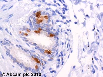
  • Immunohistochemistry (Formalin/PFA-fixed paraffin-embedded sections) – HE4 antibody (ab24480)Anti-HE4 antibodyab24480(enlarge)

Applications

Show applications key

Our Abpromise guarantee covers the use of ab24480 in the following tested applications.

The application notes include recommended starting dilutions; optimal dilutions/concentrations should be determined by the end user.

  • ShowHide

    WB

    WB: 1/100. Detects a ban…Read more →

    WB: 1/100. Detects a band of approximay 25 kDa (predicted molecular weight: 13 kDa).

  • ShowHide1 Image

    IHC-P

    IHC-P: 1/40. Perform hea…Read more →

    IHC-P: 1/40. Perform heat mediated antigen retrieval before commencing with IHC staining protocol.

Target

Tissue specificity

Expressed in a number of normal tissues, including male reproductive system, regions of the respiratory tract and nasopharynx. Highly expressed in a number of tumors cells lines, such ovarian, colon, breast, lung and renal cells lines. Initially described as being exclusively transcribed in the epididymis.

Sequence similarities

Contains 2 WAP domains.

Cellular localization

Secreted.

Target information above from: UniProt accessionQ14508The UniProt Consortium
The Universal Protein Resource (UniProt) in 2010
Nucleic Acids Res. 38:D142-D148 (2010).

Information by UniProt

Form

There are 5 isoforms produced by alternative splicing.

Alternative names

  • dJ461P17.6 antibody
  • EDDM4 antibody
  • epididymal protein 4 antibody
  • Epididymal secretory protein E4 antibody
  • Epididymis specific whey acidic protein type four disulfide core antibody
  • HE 4 antibody
  • Major epididymis specific protein E4 antibody
  • Major epididymis-specific protein E4 antibody
  • MGC57529 antibody
  • Putative protease inhibitor WAP5 antibody
  • WAP 5 antibody
  • WAP domain containing protein HE4 antibody
  • WAP four disulfide core domain 2 antibody
  • WAP four disulfide core domain protein 2 antibody
  • WAP four-disulfide core domain protein 2 antibody
  • WAP5 antibody
  • WFDC 2 antibody
  • WFDC2 antibody
  • WFDC2_HUMAN antibody

see all

Database links

  • Entrez Gene: 10406 Human
  • SwissProt: Q14508 Human
  • Unigene: 2719 Human

 详细产品信息可和选购

KPL Anti-Mouse IgM (mu) Antibody074-1803

KPL Anti-Mouse IgM (mu) Antibody

  • 产品型号:  074-1803
  • 简单描述
  • KPL Anti-Mouse IgM (mu) AntibodyHuman Serum Adsorbed and Peroxidase labeled
详细介绍

KPL Anti-Mouse IgM (mu) Antibody

Affinity purified polyclonal antibody to Mouse IgM heavy chains (mu) made in Goat and labeled with horseradish peroxidase. Product has been cross-adsorbed to human serum to minimize cross-reactivity to human immunoglobulin. Product is in lyophilized form. Each lot is tested to assure specificity and lot-to-lot consistency using KPL's in-house ELISA assay.


Anti-Bovine Osteocalcin酶试剂盒Takara


Anti-Bovine Osteocalcin
品牌 Code No. 产品名称 包装量 价格(元) 说明书 数量
Takara M041 Anti-Bovine Osteocalcin, Monoclonal (Clone OC4-30) 0.1 mg ¥3,117 Anti-Bovine Osteocalcin Anti-Bovine Osteocalcin Anti-Bovine Osteocalcin
Takara M042 Anti-Bovine Osteocalcin, Monoclonal (Clone OCG2) 0.1 mg ¥2,985 Anti-Bovine Osteocalcin Anti-Bovine Osteocalcin Anti-Bovine Osteocalcin
Takara M043 Anti-Bovine Osteocalcin, Monoclonal (Clone OCG3) 0.1 mg ¥2,985 Anti-Bovine Osteocalcin Anti-Bovine Osteocalcin Anti-Bovine Osteocalcin
Takara M044 Anti-Bovine Osteocalcin, Monoclonal (Clone OCG4) 0.1 mg ¥2,985 Anti-Bovine Osteocalcin Anti-Bovine Osteocalcin Anti-Bovine Osteocalcin
收藏产品 加入购物车
 
■ 制品内容
单克隆抗体 (冻干品) 0.1 mg
 
■ 制品说明
以牛骨来源Osteocalcin为抗原获得的小鼠单克隆抗体。
克隆编号 特异性 亚型
OC4-30 第17位的γ-羧基化的骨钙素 (不与脱碳酸骨钙素反应) Mouse IgG2a
OCG2 45-49残基 Mouse IgG1
OCG3 21-31残基 Mouse IgG3
OCG4 4-9残基 Mouse IgG1
 
■ 保存
4℃。
本制品不含防腐剂。溶解后的抗体溶液 (2.0 mg/ml) 分成小包装后,-20℃可保存1年,添加0.1%NaN3后4℃可保存6个月。避免反复冻融,稀释后尽量不要保存。
 
■ 抗体溶解
使用50 μl灭菌蒸馏水溶解 (2.0 mg/ml,不含防腐剂)。
 
■ 用途
◇ 还原和非还原条件下进行蛋白质印迹分析。
◇ 冷冻切片的免疫组织化学染色。
◇ OC4-30、OCG2、OCG4:石蜡包埋切片的免疫组织化学染色。


页面更新:2021-01-19 16:28:19

Anti-TF, Monoclonal酶试剂盒Takara


Anti-TF, Monoclonal
品牌 Code No. 产品名称 包装量 价格(元) 说明书 数量
Takara M201 Anti-TF, Monoclonal 0.1 mg ¥2,553 Anti-TF, Monoclonal Anti-TF, Monoclonal Anti-TF, Monoclonal
收藏产品 加入购物车
 
■ 制品内容
单克隆抗体 (冻干品) 0.1 mg
 
■ 制品说明
以Trigger Farctor 伴侣蛋白部分氨基酸序列(165178)-KLH复合物为抗原获得的小鼠单克隆抗体。
克隆编号 特异性 亚型
TF 19-7F  与使用pCold TF DNA表达的TF标签融合蛋白质特异性反应。 Mouse IgG1
 
■ 保存
4℃。
本制品不含防腐剂。溶解后的抗体溶液(2.0 mg/ml)分成小包装后,-20℃可保存1年,添加0.1%NaN3后4℃可保存6个月。避免反复冻融,稀释后尽量不要保存。
 
■ 抗体溶解
使用50 μl灭菌蒸馏水溶解(2.0 mg/ml,不含防腐剂)。
 
■ 用途
检测使用pCold TF DNA表达的TF标签融合蛋白质。
◇ 还原及非还原条件下的蛋白质印迹分析。
◇ 免疫沉降。
 

页面更新:2021-01-15 16:33:40

Anti-ProS2, Monoclonal酶试剂盒Takara


Anti-ProS2, Monoclonal
品牌 Code No. 产品名称 包装量 价格(元) 说明书 数量
Takara M200 Anti-ProS2, Monoclonal 0.1 mg Inquire Anti-ProS2, Monoclonal Anti-ProS2, Monoclonal Anti-ProS2, Monoclonal
收藏产品 加入购物车
 
■ 制品内容
单克隆抗体 (冻干品) 0.1 mg
 
■ 制品说明
以ProS 2* 标签抗原蛋白质为抗原的小鼠单克隆抗体。
*ProS2:粘球菌Myxococcus xanthus来源的分子量为23 kDa的可溶性标签,是可溶性蛋白Protein S 在N-terminal domain的串联二聚体。可用于大肠杆菌冷休克表达载体pCold ProS2 DNA(Code No.: 3371)
克隆编号 特异性 亚型
ProS 7B-8F
与pCold ProS2 DNA表达的ProS2标签融合蛋白质的ProS2特异性反应
Mouse IgG1
 
■ 保存
4℃。
本制品不含防腐剂。溶解后的抗体溶液(2.0 mg/ml)分成小包装后,-20℃可保存1年,添加0.1%NaN3后4℃可保存6个月。避免反复冻融,稀释后尽量不要保存。
 
■ 抗体溶解
使用50 μl灭菌蒸馏水溶解(2.0 mg/ml,不含防腐剂)。
 
■ 用途
用于检测pCold ProS2 DNA表达的ProS2 标签融合蛋白质。
◇ 还原及非还原条件下的蛋白质印迹分析。
◇ 免疫沉降。
 

页面更新:2021-01-15 16:42:34

Anti-Human iPS酶试剂盒Takara


Anti-Human iPS
品牌 Code No. 产品名称 包装量 价格(元) 说明书 数量
Takara M221 Anti-Human Oct4, Monoclonal 0.1 mg Inquire Anti-Human iPS Anti-Human iPS Anti-Human iPS
Takara M222 Anti-Human Lin28, Monoclonal 0.1 mg Inquire Anti-Human iPS Anti-Human iPS Anti-Human iPS
Takara M223 Anti-Human Sox2, Monoclonal 0.1 mg Inquire Anti-Human iPS Anti-Human iPS Anti-Human iPS
收藏产品 加入购物车
 
■ 制品内容
单克隆抗体 (冻干品) 0.1 mg
 
■ 制品说明
以基因重组Human Oct4全长蛋白质、Human Lin28全长蛋白质、Human Sox2肽 (219-236) [GSPTYSMSYSQQGTPGMA]-KLH复合物为免疫原获得的小鼠单克隆抗体。
克隆编号 特异性 亚型
Oct4 13-12F 与人Oct4特异性反应 Mouse IgG2b
Lin28 91-12A 与人Lin28特异性反应 Mouse IgG2b
Sox2 9B-12H 与人Sox2特异性反应 Mouse IgG2b
 
■ 保存
4℃。
本制品不含防腐剂。溶解后的抗体溶液 (2.0 mg/ml) 分成小包装后,-20℃可保存1年,添加0.1%NaN3后4℃可保存6个月。避免反复冻融,稀释后尽量不要保存。
 
■ 抗体溶解
使用50 μl灭菌蒸馏水溶解 (2.0 mg/ml,不含防腐剂)。
 
■ 用途
◇ 还原条件下的蛋白质印迹分析。
◇ 免疫细胞化学染色。


页面更新:2021-01-18 11:09:26

Anti-Rat Bone Specific Alkaline Phosphatase, Polyclonal酶试剂盒Takara


Anti-Rat Bone Specific Alkaline Phosphatase, Polyclonal
品牌 Code No. 产品名称 包装量 价格(元) 说明书 数量
Takara M190 Anti-Rat Bone Specific Alkaline Phosphatase, Polyclonal 0.1 mg ¥3,495 Anti-Rat Bone Specific Alkaline Phosphatase, Polyclonal Anti-Rat Bone Specific Alkaline Phosphatase, Polyclonal Anti-Rat Bone Specific Alkaline Phosphatase, Polyclonal
收藏产品 加入购物车
 
■ 制品内容
多克隆抗体 (冻干品) 0.1 mg
 
■ 制品说明
以人及大鼠骨特异性碱性磷酸酶的高同源性肽 (20-49) [PEKEKDPKYWRDQAQETLKYALELQKLNTN]-KLH (Keyhole limpet hemocyanin) 复合物为免疫原获得的兔多克隆抗体。
 
■ 保存
4℃。
本制品不含防腐剂。溶解后的抗体溶液 (2.0 mg/ml) 分成小包装后,-20℃可保存1年,添加0.1%NaN3后4℃可保存6个月。避免反复冻融,稀释后尽量不要保存。
 
■ 特异性
与大鼠骨特异性碱性磷酸酶特异性反应。
 
■ 抗体溶解
使用50 μl灭菌蒸馏水溶解 (2.0 mg/ml,不含防腐剂)。
 
■ 用途
◇ 石蜡包埋切片的免疫组织化学染色*
     * 无需抗原的修复。
◇ 非还原条件下的蛋白质印迹法对骨特异性碱性磷酸酶的检出。


页面更新:2021-01-18 11:19:21

Anti-Human E-cadherin酶试剂盒Takara


Anti-Human E-cadherin
品牌 Code No. 产品名称 包装量 价格(元) 说明书 数量
Takara M106 Anti-Human E-cadherin, Monoclonal(Clone HECD-1) 0.1 mg ¥3,505 Anti-Human E-cadherin Anti-Human E-cadherin Anti-Human E-cadherin
Takara M126 Anti-Human E-cadherin, Monoclonal(Clone SHE78-7) 0.1 mg Inquire Anti-Human E-cadherin Anti-Human E-cadherin Anti-Human E-cadherin
收藏产品 加入购物车
 
■ 制品内容
单克隆抗体 (冻干品) 0.1 mg
 
■ 制品说明
HECD-1:以人乳腺癌细胞MCF-7为抗原获得的小鼠单克隆抗体。
SHE78-7:以人胎盘来源的可溶性E-cadherin为抗原获得的小鼠单克隆抗体。
克隆编号 特异性 亚型
HECD-1 与Human E-cadherin (细胞外domain) 特异性反应;抑制E-cadherin介导的人细胞间黏附。 Mouse IgG1
SHE78-7 与Human E-cadherin特异性反应;抑制E-cadherin介导的人细胞间黏附。 Mouse IgG2a
 
■ 保存
4℃。
本制品不含防腐剂。溶解后的抗体溶液 (2.0 mg/ml) 分成小包装后,-20℃可保存1年,添加0.1%NaN3后4℃可保存6个月。避免反复冻融,稀释后尽量不要保存。
 
■ 抗体溶解
使用50 μl灭菌蒸馏水溶解 (2.0 mg/ml,不含防腐剂)。
 
■ 用途
HECD-1
◇ E-cadherin介导的细胞间黏附机制的解析。
◇ 还原及非还原条件下的蛋白质印迹法的检出。
◇ 冷冻切片和石蜡包埋切片的免疫组织化学染色。
SHE78-7
◇ E-cadherin介导的细胞间黏附机制的解析。
◇ 还原及非还原条件下的蛋白质印迹法的检出。
◇ 冷冻切片和石蜡包埋切片的免疫组织化学染色。


页面更新:2021-01-18 11:29:27

Anti-Mouse E-cadherin酶试剂盒Takara


Anti-Mouse E-cadherin
品牌 Code No. 产品名称 包装量 价格(元) 说明书 数量
Takara M107 Anti-Mouse E-cadherin, Monoclonal (Clone ECCD-1) 0.1 mg ¥2,543 Anti-Mouse E-cadherin Anti-Mouse E-cadherin Anti-Mouse E-cadherin
Takara M108 Anti-Mouse E-cadherin, Monoclonal (Clone ECCD-2) 0.1 mg ¥2,543 Anti-Mouse E-cadherin Anti-Mouse E-cadherin Anti-Mouse E-cadherin
收藏产品 加入购物车
 
■ 制品内容
单克隆抗体 (冻干品) 0.1 mg
 
■ 制品说明
ECCD-1:以小鼠畸胎癌F9细胞为抗原获得的大鼠单克隆抗体。
ECCD-2:以小鼠畸胎癌和肝脏来源的E-cadherin片断为抗原获得的大鼠单克隆抗体。
克隆编号 特异性 亚型
ECCD-1 与Mouse E-cadherin特异性反应 (细胞间黏附抑制实验用); 抑制E-cadherin介导的小鼠细胞间黏附。 Rat IgG2b
ECCD-2 与Mouse E-cadherin特异性反应 (免疫组化染色用)。 Rat IgG2a
 
■ 保存
4℃。
本制品不含防腐剂。溶解后的抗体溶液 (2.0 mg/ml) 分成小包装后,-20℃可保存1年,添加0.1%NaN3后4℃可保存6个月。避免反复冻融,稀释后尽量不要保存。
 
■ 抗体溶解
使用50 μl灭菌蒸馏水溶解 (2.0 mg/ml,不含防腐剂)。
 
■ 用途
ECCD-1:用于细胞间黏附抑制实验。
ECCD-2:
◇ 还原及非还原条件下的蛋白质印迹分析。
◇ 冷冻切片的免疫组织化学染色。


页面更新:2021-01-18 11:56:03

Anti-Human N-cadherin, Monoclonal酶试剂盒Takara


Anti-Human N-cadherin, Monoclonal
品牌 Code No. 产品名称 包装量 价格(元) 说明书 数量
Takara M180 Anti-Human N-cadherin, Monoclonal 0.1 mg ¥2,543 Anti-Human N-cadherin, Monoclonal Anti-Human N-cadherin, Monoclonal Anti-Human N-cadherin, Monoclonal
收藏产品 加入购物车
 
■ 制品内容
单克隆抗体 (冻干品) 0.1 mg
 
■ 制品说明
基因重组人N-cadherin细胞内蛋白 (氨基酸序列737-876) 免疫的BALB/c小鼠脾细胞与小鼠骨髓瘤细胞P3U1融合获得的杂交瘤细胞在BALB/c小鼠腹腔内增殖并抽取腹水制备的单克隆抗体。
克隆编号 特异性 亚型
Ncad 1-1-3 与Human N-cadherin的细胞内蛋白(氨基酸序列737-876)特异性反应;与Human E-cadherin、P- cadherin不反应。 Mouse IgG1
 
■ 保存
4℃。
本制品不含防腐剂。溶解后的抗体溶液 (2.0 mg/ml) 分成小包装后,-20℃可保存1年,添加0.1%NaN3后4℃可保存6个月。避免反复冻融,稀释后尽量不要保存。
 
■ 抗体溶解
使用50 μl灭菌蒸馏水溶解 (2.0 mg/ml,不含防腐剂)。
 
■ 用途
◇ 还原及非还原条件下的蛋白质印迹分析。
◇ 石蜡包埋切片和冷冻切片的免疫组织化学染色。
◇ 免疫沉降。


页面更新:2021-01-18 14:32:56

Anti-N-cadherin, Polyclonal酶试剂盒Takara


Anti-N-cadherin, Polyclonal
品牌 Code No. 产品名称 包装量 价格(元) 说明书 数量
Takara M142 Anti-N-cadherin, Polyclonal 0.4 mg ¥2,193 Anti-N-cadherin, Polyclonal Anti-N-cadherin, Polyclonal Anti-N-cadherin, Polyclonal
收藏产品 加入购物车
 
■ 制品内容
多克隆抗体 (冻干品) 0.4 mg
 
■ 制品说明
以人N-cadherin的部分氨基酸序列 (808-827) -KLH复合物为免疫原获得的兔多克隆抗体。
 
■ 保存
4℃。
本制品不含防腐剂。溶解后的抗体溶液 (2.0 mg/ml) 分成小包装后,-20℃可保存1年,添加0.1%NaN3后4℃可保存6个月。避免反复冻融,稀释后尽量不要保存。
 
特异性
与Human N-cadherin反应;不能抑制N-cadherin介导的细胞间黏附。
 
■ 抗体溶解
使用200 μl灭菌蒸馏水溶解 (2.0 mg/ml,不含防腐剂)。
 
■ 用途
◇ 石蜡包埋切片和冷冻切片的免疫组织化学染色。
◇ 细胞组织提取液在非还原非加热条件下的蛋白质印迹分析。


页面更新:2021-01-18 14:36:35

Anti-Mouse P-cadherin, Monoclonal (Clone PCD-1)酶试剂盒Takara


Anti-Mouse P-cadherin, Monoclonal (Clone PCD-1)
品牌 Code No. 产品名称 包装量 价格(元) 说明书 数量
Takara M109 Anti-Mouse P-cadherin, Monoclonal (Clone PCD-1) 0.1 mg ¥3,505 Anti-Mouse P-cadherin, Monoclonal (Clone PCD-1) Anti-Mouse P-cadherin, Monoclonal (Clone PCD-1) Anti-Mouse P-cadherin, Monoclonal (Clone PCD-1)
收藏产品 加入购物车
 
■ 制品内容
单克隆抗体 (冻干品) 0.1 mg
 
■ 制品说明
以小鼠endoderm cell line PSA5-E为抗原获得的大鼠单克隆抗体。
克隆编号 特异性 亚型
PCD-1 与MouseP-cadherin特异性反应;抑制P-cadherin介导的小鼠细胞间黏附。 Rat IgG2a
 
■ 保存
4℃。
本制品不含防腐剂。溶解后的抗体溶液 (2.0 mg/ml) 分成小包装后,-20℃可保存1年,添加0.1%NaN3后4℃可保存6个月。避免反复冻融,稀释后尽量不要保存。
 
■ 抗体溶解
使用50 μl灭菌蒸馏水溶解 (2.0 mg/ml,不含防腐剂)。
 
■ 用途
◇ P-cadherin介导的细胞连接机制的解析。
◇ 还原及非还原条件下的蛋白质印迹分析。
◇ 冷冻切片的免疫组织化学染色。
◇ 基因重组体P-cadherin的解析。


页面更新:2019-07-10 10:58:37

Anti-Dentin Matrix Protein1, Polyclonal酶试剂盒Takara


Anti-Dentin Matrix Protein1, Polyclonal
品牌 Code No. 产品名称 包装量 价格(元) 说明书 数量
Takara M176 Anti-Dentin Matrix Protein1, Polyclonal 0.1 mg ¥3,275 Anti-Dentin Matrix Protein1, Polyclonal Anti-Dentin Matrix Protein1, Polyclonal Anti-Dentin Matrix Protein1, Polyclonal
收藏产品 加入购物车
 
■ 制品内容
多克隆抗体 (冻干品) 0.1 mg
 
■ 制品说明
以大鼠Dentin Matrix Protein-1 (DMP-1) N末端附近的氨基酸序列 (90-111)-KLH复合物为免疫原获得的兔多克隆抗体。
 
■ 保存
4℃。
本制品不含防腐剂。溶解后的抗体溶液 (2.0 mg/ml) 分成小包装后,-20℃可保存1年,添加0.1%NaN3后4℃可保存6个月。避免反复冻融,稀释后尽量不要保存。
 
特异性
与大鼠DMP-1特异性反应。
 
■ 抗体溶解
使用50 μl灭菌蒸馏水溶解 (2.0 mg/ml,不含防腐剂)。
 
■ 用途
◇ 还原及非还原条件下的蛋白质印迹分析。
◇ 石蜡包埋切片的免疫组织化学染色。


页面更新:2021-01-18 16:17:55

Anti-Human Fibronectin酶试剂盒Takara


Anti-Human Fibronectin
品牌 Code No. 产品名称 包装量 价格(元) 说明书 数量
Takara M002 Anti-Human Fibronectin, Monoclonal (Clone FN12-8) 0.4 mg ¥2,634 Anti-Human Fibronectin Anti-Human Fibronectin Anti-Human Fibronectin
Takara M010 Anti-Human Fibronectin, Monoclonal (Clone FN30-8) 0.4 mg ¥2,634 Anti-Human Fibronectin Anti-Human Fibronectin Anti-Human Fibronectin
收藏产品 加入购物车
 
■ 制品内容
单克隆抗体 (冻干品) 0.4 mg
 
■ 制品说明
以人血浆Fibronectin抗原获得的小鼠单克隆抗体。
克隆编号 特异性 亚型
Clone FN12-8 细胞结合域 Mouse IgG1
Clone FN30-8 细胞结合域 Mouse IgG1
 
■ 保存
4℃。
本制品不含防腐剂。溶解后的抗体溶液 (2.0 mg/ml) 分成小包装后,-20℃可保存1年,添加0.1%NaN3后4℃可保存6个月。避免反复冻融,稀释后尽量不要保存。
 
■ 抗体溶解
使用200 μl灭菌蒸馏水溶解 (2.0 mg/ml,不含防腐剂)。
 
■ 用途
◇ 非还原非加热条件下的蛋白质印迹分析。
◇ 石蜡包埋切片及冷冻切片的免疫组织化学染色。
◇ 黏附抑制实验。


页面更新:2021-01-18 16:24:53

Anti-Glucagon, Polyclonal酶试剂盒Takara


Anti-Glucagon, Polyclonal
品牌 Code No. 产品名称 包装量 价格(元) 说明书 数量
Takara M182 Anti-Glucagon, Polyclonal 0.1 mg ¥2,864 Anti-Glucagon, Polyclonal Anti-Glucagon, Polyclonal Anti-Glucagon, Polyclonal
收藏产品 加入购物车
 
■ 制品内容
多克隆抗体 (冻干品) 0.1 mg
 
■ 制品说明
以人胰高血糖素 (Glucagon) [HSQGTFTSDYSKYLDSRRAQDFVQWLMNT]-KLH复合物为免疫原后获得的豚鼠多克隆抗体。
 
■ 保存
4℃。
本制品不含防腐剂。溶解后的抗体溶液 (2.0 mg/ml) 分成小包装后,-20℃可保存1年,添加0.1%NaN3后4℃可保存6个月。避免反复冻融,稀释后尽量不要保存。
 
特异性
可识别人胰高血糖素 (Glucagon) 氨基酸序列:HSQGTFTSDYSKYLDSRRAQDFVQWLMNT
 
■ 抗体溶解
使用50 μl灭菌蒸馏水溶解 (2.0 mg/ml,不含防腐剂)。
 
■ 用途
石蜡包埋切片和冷冻切片的免疫组织化学染色。


页面更新:2021-01-19 11:51:34

Anti-Heme Oxygenase-1酶试剂盒Takara


Anti-Heme Oxygenase-1
品牌 Code No. 产品名称 包装量 价格(元) 说明书 数量
Takara M174 Anti-Heme Oxygenase-1 (GTS-1), Monoclonal 0.1 mg Inquire Anti-Heme Oxygenase-1 Anti-Heme Oxygenase-1 Anti-Heme Oxygenase-1
Takara M175 Anti-Heme Oxygenase-1 (GTS-3), Monoclonal 0.1 mg ¥3,026 Anti-Heme Oxygenase-1 Anti-Heme Oxygenase-1 Anti-Heme Oxygenase-1
收藏产品 加入购物车
 
■ 制品内容
单克隆抗体 (冻干品) 0.1 mg
 
■ 制品说明
以大鼠Heme Oxygenase-1为抗原的小鼠单克隆抗体。
克隆编号 特异性 亚型
GTS-1 与Human和Rat Heme Oxygenase-1特异性反应;抑制人和大鼠Heme Oxygenase-1酶活性。 Mouse IgG1
GTS-3 Mouse IgG1
 
■ 保存
4℃。
本制品不含防腐剂。溶解后的抗体溶液 (2.0 mg/ml) 分成小包装后,-20℃可保存1年,添加0.1%NaN3后4℃可保存6个月。避免反复冻融,稀释后尽量不要保存。
 
■ 抗体溶解
使用50 μl灭菌蒸馏水溶解 (2.0 mg/ml,不含防腐剂)。
 
■ 用途
◇ 蛋白质印迹法检出血红素加氧酶。
◇ 石蜡包埋切片及冰冻切片的免疫组织化学染色。
◇ 血红素加氧酶活性抑制实验。
◇ 人源组织的石蜡包埋切片使用GTS-1。
◇ 大鼠组织的石蜡包埋切片使用GTS-3。


页面更新:2021-01-19 16:00:44

Anti-Human Influenza Virus酶试剂盒Takara


Anti-Human Influenza Virus
品牌 Code No. 产品名称 包装量 价格(元) 说明书 数量
Takara M145 Anti-Human Influenza A (H1N1, H2N2), Monoclonal (Clone C179) 0.1 mg ¥2,353 Anti-Human Influenza Virus Anti-Human Influenza Virus Anti-Human Influenza Virus
Takara M146 Anti-Human Influenza A (H3N2), Monoclonal (Clone F49) 0.1 mg ¥2,353 Anti-Human Influenza Virus Anti-Human Influenza Virus Anti-Human Influenza Virus
Takara M147 Anti-Human Influenza A (H1, H2, H3), Monoclonal (Clone C111) 0.1 mg ¥2,353 Anti-Human Influenza Virus Anti-Human Influenza Virus Anti-Human Influenza Virus
Takara M148 Anti-Human Influenza B, Monoclonal (Clone 9D6) 0.1 mg ¥2,423 Anti-Human Influenza Virus Anti-Human Influenza Virus Anti-Human Influenza Virus
收藏产品 加入购物车
 
■ 制品内容
单克隆抗体 (冻干品) 0.1 mg
 
■ 制品说明
流感病毒抗原的小鼠单克隆抗体
C179
识别甲型流感病毒H1N1及H2N2的HA干区保守序列立体结构的抗体。H1亚型和H2亚型的反应不受抗原变异的影响,且对于这两种亚型具有较强的中和活性。
F49
特异性识别甲型流感病毒 (H3N2) 型HA干区的抗体,没有中和活性。
C111
识别甲型流感病毒Matrix Protein (M1) 的抗体。与甲型 (A型) 病毒所有的亚型都可以反应。本抗体可有效用于细胞染色、免疫沉淀、蛋白质印迹等。
9D6
识别乙型流感病毒核蛋白质 (NP) 的抗体 。可与广泛范围的乙型流感病毒特异性反应。可用于细胞染色、免疫沉淀、蛋白质印迹等。
克隆编号 特异性 亚型
C179 ◇ 与甲型流感病毒H1N1及H2N2亚型的HA蛋白分子氨基酸序列的结构保守区特异性反应,不与甲型流感H3N2亚型的HA蛋白分子交叉反应。
◇ 具有中和H1N1及H2N2亚型病毒的活性。
◇ 已确认可与禽流感H5N3反应。
◇ 对所有亚型病毒的血细胞凝集抑制试验没有任何影响。
Mouse IgG2a
F49 ◇ 与甲型流感H3N2亚型的HA蛋白分子氨基酸序列的结构保守区特异性反应,不与甲型流感H1N1及H2N2亚型的HA蛋白分子交叉反应。
◇ 不具有中和流感病毒的活性。
◇ 除人流感病毒外,还可与A/duck/Czechoslovakia/1/56 (H4N6)、A/ chiken/Germany"N"/49 (H10N7)交叉反应。
◇ 对所有亚型病毒的血细胞凝集抑制试验没有任何影响。
Mouse IgG1
C111 ◇ 与甲型流感H1N1、H2N2、H3N2亚型的Matrix Protein特异性反应,不与乙型流感交叉反应。 Mouse IgG2a
9D6 ◇ 与乙型流感病毒的核蛋白质 (NP) 特异性反应,不与甲型流感的所有亚型交叉反应。 Mouse IgG1
 
■ 保存
4℃。
本制品不含防腐剂。溶解后的抗体溶液 (2.0 mg/ml) 分成小包装后,-20℃可保存1年,添加0.1%NaN3后4℃可保存6个月。避免反复冻融,稀释后尽量不要保存。
 
■ 抗体溶解
使用50 μl灭菌蒸馏水溶解 (2.0 mg/ml,不含防腐剂)。
 
■ 用途
流感病毒的检测和分型。
C179:染色实验 (PAP法);流感病毒H1N1与H2N2的中和实验;实验动物对流感病毒感染的预防效果。
F49:染色实验 (PAP法)。
C111:染色实验 (PAP法);蛋白质印迹。
9D6:染色实验 (PAP法);蛋白质印迹。


页面更新:2021-01-19 16:09:08