Phusion® 超保真 PCR 预混液(提供 HF 缓冲液) 货 号 M0531LNEB酶试剂 New England Biolabs

上海金畔生物科技有限公司代理New England Biolabs(NEB)酶试剂全线产品,欢迎访问官网了解更多产品信息和订购。

产品资料 – DNA聚合酶与扩增技术 – 高保真PCR

Phusion® 超保真 PCR 预混液(提供 HF 缓冲液)                              收藏

Phusion® 超保真 PCR 预混液(提供 HF 缓冲液)                 货   号                  M0531L Phusion® 超保真 PCR 预混液(提供 HF 缓冲液)                 货   号                  M0531L Phusion® 超保真 PCR 预混液(提供 HF 缓冲液)                 货   号                  M0531L Phusion® 超保真 PCR 预混液(提供 HF 缓冲液)                 货   号                  M0531L Phusion® 超保真 PCR 预混液(提供 HF 缓冲液)                 货   号                  M0531L

货 号
规 格
价 格(元)
北京库存
上海库存
广州库存
成都库存
苏州库存

#M0531L
500 次反应(50 μl 反应体系)
6,789.00

#M0531S
100 次反应(50 μl 反应体系)
1,759.00

Download:       

  • isoschizomers     |
  • compatible ends     | 
  • single letter code

相关产品

Phusion® 超保真 DNA 聚合酶

Phusion® 超保真 PCR 预混液( 提供 HF 缓冲液 )

Phusion® 超保真 PCR 预混液( 提供 GC 缓冲液 )

Phusion® 热启动 Flex DNA 聚合酶

Phusion® 热启动 Flex 2X 预混液

Phusion® 超保真 PCR 试剂盒

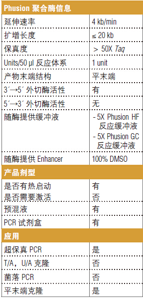
Phusion 聚合酶信息

Phusion® 超保真 PCR 预混液(提供 HF 缓冲液)                 货   号                  M0531L

概述

由于 DNA 序列需要在扩增后进行校正,所以拥有高保真度的 DNA 聚合酶对于 PCR 应用至关重要。由 NEB 生产、质量控制的 Thermo Scientific® Phusion 超保真 DNA 聚合酶兼备高保真度和稳定扩增的性能,适用于所有 PCR 应用。其独特的结构包括一个类似 Pyrococcus 的酶和一个掺入率增强结构域,二者融合在一起。这种组合提升了聚合反应的保真性和速度。可供选择的试剂包括单酶、预混液和试剂盒。Phusion 热启动 Flex DNA 聚合酶有单酶和预混液剂型可供选择。Phusion DNA 聚合酶可用于长片段的扩增,是克隆实验的理想
选择。

其它剂型

Phusion 和 Phusion 热启动 Flex DNA 聚合酶均有预混液剂型可供选择,Phusion 预混液有含 HF 或 GC 缓冲液两种形式。
Phusion PCR 试剂盒包含 Phusion 聚合酶、Phusion HF 和 GC 缓冲液、dNTPs、MgCl2、DMSO 和 DNA
Marker。

浓度

2,000 units/ml。

Phusion® 超保真 PCR 预混液(提供 HF 缓冲液)                 货   号                  M0531L
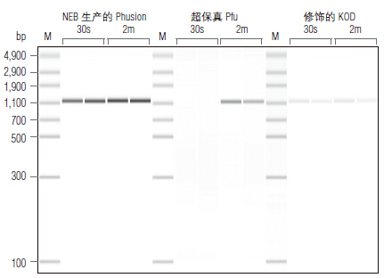
Phusion 超保真 DNA 聚合酶扩增片段的产量更高,延伸时间更短。使用不同的聚合酶扩增一段 1.2 kb 长
度的 C. elegans 基因组片段。所有扩增反应按照厂家推荐条件重复进行(30 个循环、30 秒或 2 分钟的延伸时间)并用微流控芯片技术(microfluidic LabChip® analysis)检测。

Phusion® 超保真 PCR 预混液(提供 HF 缓冲液)                 货   号                  M0531L
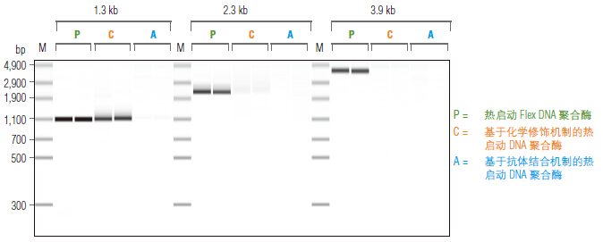
Phusion 热启动 Flex DNA 聚合酶具有超强扩增能力。
除了 1.3 kb C. elegans 扩增片段,其它扩增片
段模板均为 human Jurkat。扩增长度如上胶图所示。所有扩增反应按照厂家推荐条件(30 个热循
环)重复进行并用微流控芯片技术(microfluidic LabChip® analysis)检测。

* Phusion DNA Polymerase was developed by Finnzymes Oy, now a part of Thermo Fisher Scientific.
This product is manufactured by New England Biolabs, Inc. under agreement with, and under the performance specifications of Thermo Fisher Scientific. Phusion® is a registered trademark and property of Thermo Fisher Scientific.

许可/专利/免责:参见 390-391 页。

Phusion® 超保真 PCR 预混液(提供 HF 缓冲液)                 货   号                  M0531L

日本同仁化学Hampton蛋白结晶试剂盒Glycerol/HR2-623- DOJINDO

上海金畔生物科技有限公司代理日本同仁化学 DOJINDO代理商全线产品,欢迎访问官网了解更多信息

上海金畔生物代理Hampton research品牌蛋白结晶试剂耗材工具等,我们将竭诚为您服务,欢迎访问Hampton research官网或者咨询我们获取更多相关Hampton research品牌产品信息。

Products > Optimize Reagents > Optimize – Organics (Non-Volatile) > Glycerol

Glycerol

Hampton蛋白结晶试剂盒Glycerol/HR2-623


CAT NO

HR2-623

NAME

100% Glycerol

DESCRIPTION

100 mL

PRICE

$84.00

支持材料

Hampton蛋白结晶试剂盒Glycerol/HR2-623 HR2-623 Glycerol SDS

应用

  • Crystallization grade Glycerol for formulating screens, optimization and cryoprotection
  • 结晶级甘油,用于配制筛选、优化和冷冻保护

特征

  • Gradually increasing the Glycerol concentration to 5 to 35% in the original crystallization reagent is often times an effective cryoprotectant.
  • 将原始结晶试剂中的甘油浓度逐渐增加到 5% 到 35% 通常是一种有效的冷冻保护剂。

描述Glycerol

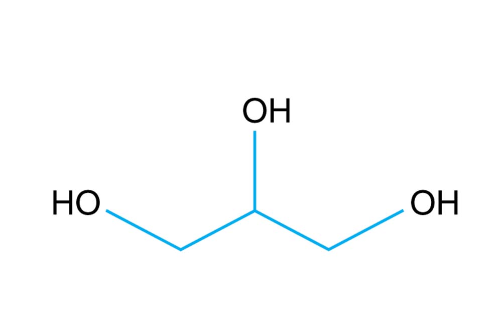
Synonyms: 1,2,3-Propanetriol or Glycerin
HOCH2CH(OH)CH2OH
C3H8O3
Mr 92.09
CAS Number [56-81-5]
EC Number 200-289-5
Beilstein Registry Number 635685
RTECS MA8050000
MDL Number MFCD00004722
PubChem Substance ID 24895252
Purity ≥ 99.0% (GC)
pKa 14.2

Measured pH range of 100% Glycerol: 5.0 – 7.7 at 25°C
Measured Conductivity Range: 0.0 – 0.2 µS/cm at 25°C
Measured Refractive Index Range: 1.47124 – 1.47450 at 20°C

同义词:1,2,3-丙三醇或甘油
HOCH2CH(OH)CH2OH
C3H8O3
92.09 先生
CAS 编号 [56-81-5]
欧盟编号 200-289-5
贝尔斯坦注册号 635685
RTECS MA8050000
MDL 编号 MFCD00004722
PubChem 物质 ID 24895252
纯度 ≥ 99.0% (GC)
pKa 14.2

测得的 100% 甘油的 pH 值范围:25°C 时为 5.0 – 7.7
测量电导率范围:25°C 时 0.0 – 0.2 µS/cm
测得的折射率范围:20°C 时为 1.47124 – 1.47450

Al  ≤0.0005%
Ca ≤0.0005%
Cd ≤0.0001%
Cl  ≤0.001%
Cu ≤0.0005%
Fe ≤0.0005%
K   ≤0.005%
Mg ≤0.0005%
NH3 ≤0.05%
NH4+ ≤0.05%
Na ≤0.005%
P  ≤0.0005%
Pb ≤0.001%
SO4 ≤0.002%
Zn ≤0.0005%

参考Glycerol

1. Crystallization and Preliminary X-ray Analysis of the Auto-inhibited Twitchin Kinase. Hu, S-H; Lei, JY; Wilce, MCJ; Valenzuela, MRL; Benian, GM; Parker, MW; Kemp, BE. J Mol Biol 236, 1259- 1261, 1994.

1. 自抑制 Twitchin 激酶的结晶和初步 X 射线分析。 胡,S-H; 雷,JY; 威尔斯,MCJ; 瓦伦苏埃拉,MRL; 贝尼安,总经理; 帕克,兆瓦; 坎普,BE。 分子生物学杂志 236, 1259-1261, 1994。

PALL Acro37 TF通气过滤器4464现货供应,颇尔通气过滤器4464

PALL Acro37 TF通气过滤器4464现货供应,颇尔通气过滤器4464

适于小体积通气、排气、气体过滤的多用途滤器

PALL Acro37 TF通气过滤器4464现货供应,颇尔通气过滤器4464特点:

1.一次性滤器;与可换膜不锈钢滤器相比节省人力物力。

2.PTFE膜片和聚丙烯外壳使其具有极好的化学兼容性。

3.可高温高压灭菌。

4.经济;可提供大包装。

特性:

制造材料

过滤介质:PTFE带聚丙烯支撑层

外壳:聚丙烯

有效过滤面积:7.5cm2

zui大操作温度:100℃

zui小水透压力:30psi

标准气体流速:8Lpm at 3psi

消毒方法:非灭菌包装;可高温高压灭菌。

应用:

1.用于小体积的通风、排气

2.亦可用于小体积的溶剂过滤

订购信息- Acro37TF通气过滤器

产品编号

产品说明

包装

4464

0.2μm,37mm

24/pkg

4465

0.2μm,37mm

200/pkg

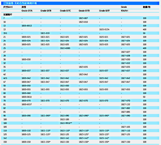
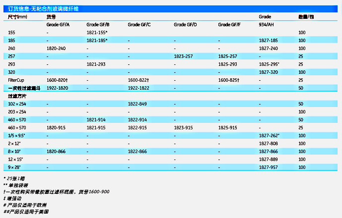
Whatman10463862无粘合剂玻璃微纤维滤纸UNIFLO 13/0.45 RC 500/PK

【简单介绍】

Whatman为客户提供多种多样的特殊实验产品,以满足实验者的不同实验需求。秉承Whatman传统的质量,这些产品集合了易于操作、精确度高、一致性好等优点。

【简单介绍】

Whatman为客户提供多种多样的特殊实验产品,以满足实验者的不同实验需求。秉承Whatman传统的质量,这些产品集合了易于操作、精确度高、一致性好等优点。

【详细说明】

原装进口英国Whatman10463862无粘合剂玻璃微纤维滤纸UNIFLO 13/0.45 RC 500/PK

英国Whatman10463862无粘合剂玻璃微纤维滤纸UNIFLO 13/0.45 RC 500/PK

产品介绍:

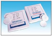
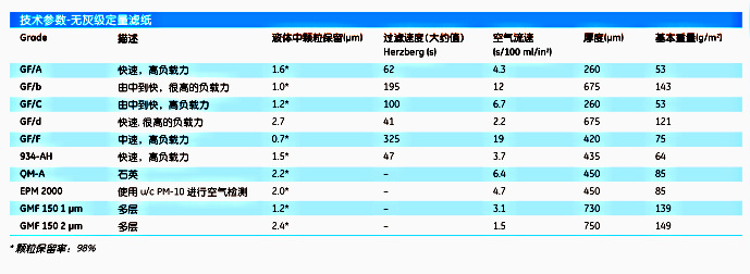
Galss Microfiber Filters玻璃微纤维滤纸

Whatman玻璃微纤维滤纸采用100%硼硅酸玻璃纤维制造而

成,包括不含粘合剂呈化学惰性的和含黏合剂的2种类型。

这种深度滤纸流速快、负载力大、保留颗粒极为细小,

可达亚微颗粒范围。玻璃微纤维滤纸能承受高达500℃的温

度,所以能用于需要灼烧的比重分析和高温气体过滤。

Whatman玻璃微纤维滤纸具有毛细纤维结构,能吸附比同

等纤维素滤纸更多的水分,使其能适用于点样分析和液闪计

数方法,并能制成全透明,用于后续的显微检验。

Whatman玻璃微纤维滤纸如GF/BGF/D具有高流速、低抗

性和高颗粒负载力的特征,是理想的预滤产品,能明显地增

加过滤量。多级GMF 150滤纸特别适合于预滤那些难过滤的

大容量溶液。

英国Whatman10463862无粘合剂玻璃微纤维滤纸UNIFLO 13/0.45 RC 500/PK

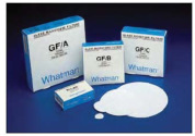
    玻璃微纤维GF系列

玻璃微纤维GF系列

无黏合剂玻璃微纤维滤纸

Grade GF/A : 1.6 μm

保留细小颗粒、流速快、负载力高,用于高效常规过滤,包

括废水水污染检测,用于过滤水、藻类和细菌培养、食品分

析、蛋白过滤和弱?发射器的放免测定,推荐用于空气污染

监测中的颗粒物重量测定、烟囱取样和吸附等。

此类滤纸也以滤杯和一次性过滤漏斗形式提供。

Grade GF/B : 1.0 μm

厚度是GF/A3倍,湿强更高,负载力也明显增强。用于保

留细小颗粒,流速快。特别适用于在液体澄清或颗粒定量中

悬浮细颗粒高的样品处理。可用于膜过滤前的细颗粒物预过

滤,用于要求高负载力的LSC技术。

Grade GF/C : 1.2 μm

保留细小颗粒,有很好的流速。在世界许多地方,这是用于

收集饮用水和天然、工业废水中的悬浮物的标准滤纸。

快速而有效地澄清含小到中等细小颗粒的水溶液。当需要更

高负载的时候,广泛地用于细胞培养、液闪计数和结合分析

方面。

此种滤纸也可用于滤杯。

Grade 934-AH : 1.5 μm

用这种普遍的滤纸截流细小颗粒的优越性,在于快流速和高

负载力下有着非常高的保留效率。这是种表面光滑,高保留

力的硼硅酸微纤维滤纸,可耐高温达500℃。指定用于测定

水中悬浮物总数去除浑浊物和细菌培养的过滤。Grade 934-

AH用于很多范围的实验室应用中。推荐用于水污染监测、

细胞培养、液闪计数和空气污染监测。

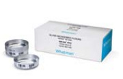
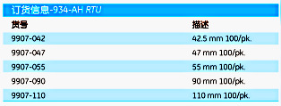
Grade 934-AH RTUNEW

Grade 934-AH,广泛用于总悬浮颗粒分析,现在为您带来全

新的Ready-To-Use (RTU)规格,帮你节省更多的时间。

934-AH RTU不含有粘合剂,由高纯的硼硅酸玻璃微纤维制成,

完全符合标准方法的要求。预洗脱和预称量的规格,无需您

做预处理,使您的试验流程更流畅。每张RTU滤纸被放置于

一个铝盘中,在铝盘边上贴的抗热标签上清晰标注了滤纸的

重量。

特征和优点

l 快速方便:可直接使用预洗、预称量的滤纸,无需再预处理

l 高效:高流速下超细颗粒保留

l 高载量:可处理非常浑浊的样品

应用

934-AH RTU符合最新版水中总悬浮颗粒的检测标准方法

2540D的要求,被广泛用于水体检测,包括:

l 河流、湖泊和海岸水体监测

l 废水处理厂的废水纯化

l 工厂排水监控

英国Whatman10463862无粘合剂玻璃微纤维滤纸UNIFLO 13/0.45 RC 500/PK


英国Whatman10463862无粘合剂玻璃微纤维滤纸UNIFLO 13/0.45 RC 500/PK


石英滤纸-Grade QM-A : 2.2μm

高纯度石英(SiO2)微纤维滤纸,特别推荐用于烟囱、烟道、

烟雾方面空气污染监测工作,操作温度可达500℃。滤纸经

热处理,无黏合剂,重金属和碱土建树含量特别低。用于

PM-10检测。QM-A根据EPA标准编号,适合大部分应用。订

货信息请参考空气样品滤纸/石英滤纸部分。

Grade GF/D : 2.7 μm

流速相当快,在相同颗粒保留度的条件下,整个过滤速度比

纤维素更快。滤纸较厚,具有较强的负载力,可作为膜的预

滤。由不同大小来适合大多数滤器。GF/D为保留细小颗粒的

膜提供了很好的保护,可以和GF/B一起使用,给膜提供了非

常有效的预滤保护。

GF/F : 0.7 μm

小到0.7μm的细小颗粒的保留率极高,不同于滤膜,它有非

常快的流速,负载力极高。

因具有严格的0.6μm-0.8μm颗粒保留度和纯硼硅酸结构,

EPA TCLP 1311毒性滤取方法是以GF/F为基质创建的。

这种滤纸在今天仍是很好的选择。推荐用于DNA吸附和纯

化,过滤细小沉淀蛋白非常有效,GF/FGF/D一起可以作

为极其难以澄清的生化溶液、流体和核酸的预滤膜。

这种滤纸同时也提供滤杯和一次性漏斗形式。

Grade EPM 2000 : 2.0 μm

EPM 2000被专门开发用来在高流量空气采样设备来收集大

气中颗粒剂气溶胶。由100%高纯度硼硅玻纤制成,用于痕

迹污染物在最低干扰和背景情况下的化学分析。

Grade GMF 150 : 1μm2μm

Whatman GMF150是一种多层玻璃微纤维滤膜,最上层是个

粗滤层(10μm),下层是孔径为1μm或者2μm的有网孔的细

滤层。采用100%硼硅酸玻璃纤维制成,没有任何添加剂,

通过很好的预滤作用来增强对颗粒的负载能力和加快过滤

速度。

英国Whatman10463862无粘合剂玻璃微纤维滤纸UNIFLO 13/0.45 RC 500/PK

英国Whatman10463862无粘合剂玻璃微纤维滤纸UNIFLO 13/0.45 RC 500/PK

英国Whatman10463862无粘合剂玻璃微纤维滤纸UNIFLO 13/0.45 RC 500/PK

订货信息:

CATALOG NO货号

DESCRIPTION描述

10463823

UNIFLO 13/0.45 PTFE 1000/PK

10463852

UNIFLO 13/0.2 RC 500/PK

10463862

UNIFLO 13/0.45 RC 500/PK

10463872

UNIFLO 13/0.2 NYL 1000/PK

10463875

UNIFLO 13/0.2 RC 1000/PK

10463876

UNIFLO 13/0.45 RC 1000/PK

10463877

UNIFLO 13/0.2 NYL 500/PK

10463878

UNIFLO 13/0.45 NYL 1000/PK

10463879

UNIFLO 13/0.45 NYL 500/PK

10463880

UNIFLO 13/0.45 PTFE 500/PK

10463881

UNIFLO 13/0.2 PTFE 1000/PK

10463882

UNIFLO 13/0.2 PTFE 500/PK


上海金畔生物科技有限公司

文章号:20783219-20783219

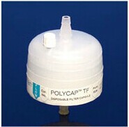
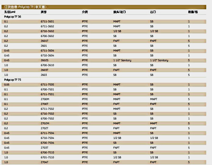
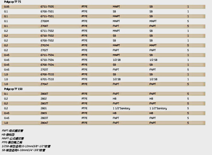
Whatman6711-3602Polycap TF囊式过滤器POLYCAP 36 0.2 PTFE 1/PK F/A

【简单介绍】

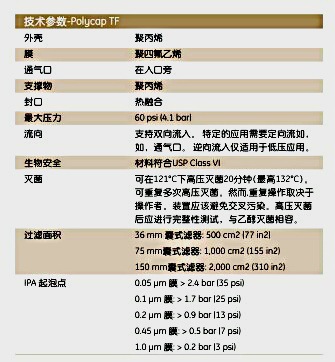
Polycap TF滤器是一次性精细囊式过滤器中的一种。这些囊

式过滤器由耐用、疏水性的PTFE膜和聚丙烯外壳制成,用于

有机和化学腐蚀性强溶液的过滤。

【简单介绍】

Polycap TF滤器是一次性精细囊式过滤器中的一种。这些囊

式过滤器由耐用、疏水性的PTFE膜和聚丙烯外壳制成,用于

有机和化学腐蚀性强溶液的过滤。

【详细说明】

原装进口英国Whatman6711-3602Polycap TF囊式过滤器POLYCAP 36 0.2 PTFE 1/PK F/A

英国Whatman6711-3602Polycap TF囊式过滤器POLYCAP 36 0.2 PTFE 1/PK F/A

产品介绍:

Polycap TF

Polycap TF滤器是一次性精细囊式过滤器中的一种。这些囊

式过滤器由耐用、疏水性的PTFE膜和聚丙烯外壳制成,用于

有机和化学腐蚀性强溶液的过滤。

特性和优点

l 对大多数溶剂有相容性,可加压加热,可测试完整性。

l 0.050.10.20.451.0μm孔径。

l 0.05μm囊式用于超滤应用;1.0μm用于延长寿命和过滤高

污染溶液。

l 可用蒸汽或乙醇加压加热消毒。

l ISO质量体系下在10000级的清洁室内环境下生产。

应用

l 通气口

l 在线过滤

l 分离

l 电子

l 制药

l 生物技术

l 实验室

l 其他用途

英国Whatman6711-3602Polycap TF囊式过滤器POLYCAP 36 0.2 PTFE 1/PK F/A

英国Whatman6711-3602Polycap TF囊式过滤器POLYCAP 36 0.2 PTFE 1/PK F/A

英国Whatman6711-3602Polycap TF囊式过滤器POLYCAP 36 0.2 PTFE 1/PK F/A

英国Whatman6711-3602Polycap TF囊式过滤器POLYCAP 36 0.2 PTFE 1/PK F/A

上海金畔生物科技有限公司

文章号19774422-19774422

thermo F1单道可调移液器 雷勃Finnpipette原装进口4641080 200ul-赛默飞中国代理商

产品信息
产品名称:
thermo F1单道可调移液器 雷勃Finnpipette原装进口4641080 200ul
产品型号:
thermo F1单道可调移液器 雷勃Finnpipette原装进口4641080 200ul 产品特点
  Finnpipette F1 0.2-2 µl micro单道可变量程移液器, CE认证 Finnpipette F1 0.2-2 µl micro, CE marked Thermo Scientific™ Finnpipette™ F1 移液器 Finnpipette F1 具有单道及多道的各种量程可供选择。其中,单道移液器具有可调和固定量程可供选择;而多道移液器可提供 8 道、12 道和 1

thermo F1单道可调移液器 雷勃Finnpipette原装进口4641080 200ul
产品详细信息:

Thermo Scientific™ Finnpipette™ F1 移液器

Finnpipette F1 具有单道及多道的各种量程可供选择。其中,单道移液器具有可调和固定量程可供选择;而多道移液器可提供 8 道、12 道和 16 道选择,其中 16 道移液器尤其适用于 384 孔样品转移。

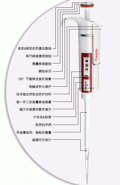
● *的液量联动装置(AVG),实现更高的精准性
● 柔软有弹性的外壳
● 轻巧的移液操作
● 120° 旋转放松指靠
● *纳米银生物安全防护
● 巧妙的设定 & 锁定防遗忘旋钮
● 超大清晰的显示屏,便于轻松设定液量
● 设计的轻触吸头推杆,将 Finnpipette 推出吸头的力量降至zui低

Thermo Scientific Finnpipette F1 单道移液器
Finnpipette F1 移液器提供zui全的从 0.2 μL 到 10 mL 的广泛液量选择,可满足您实验室手动移液的所有需求。独特的轻巧设计和抑菌材料结构使您的移液更舒适更健康,同时防遗忘按钮以及超强吹出等特点能确保您极高的移液精准性。10 μL、20 μL、50 μL 移液器各有两种吸头选择方案,能根据您实验室的吸头使用习惯选择zui合适的移液器。

thermo F1单道可调移液器 雷勃Finnpipette原装进口4641080 200ul

细胞凋亡试剂盒(PE和7-ADD标记)PE Annexin V Apoptosis Detectio

 Technical Data Sheet

PE Annexin V Apoptosis Detection Kit I

Product Information

Material Number: 559763

Component: 51-66121E

Description: 10X Annexin V Binding Buffer

Size: 50 ml (1 ea)

Storage Buffer: Aqueous buffered solution containing no preservative.

Component: 51-68981E

Description: 7-AAD

Size: 2.0 ml (1 ea)

Vol. per Test: 5 μl

Storage Buffer: Aqueous buffered solution containing fetal bovine serum and ≤0.09% sodium

azide.

Component: 51-65875X

Description: PE Annexin V

Size: 0.5 ml (1 ea)

Vol. per Test: 5 μl

Storage Buffer: Aqueous buffered solution containing BSA and ≤0.09% sodium azide.

Description

Apoptosis is a normal physiologic process which occurs during embryonic development as well as in maintenence of tissue homeostasis. The

apoptotic program is characterized by certain morphologic features, including loss of plasma membrane asymmetry and attachment,

condensation of the cytoplasm and nucleus, and internucleosomal cleavage of DNA. Loss of plasma membrane is one of the earliest features.

In apoptotic cells, the membrane phospholipid phosphatidylserine (PS) is translocated from the inner to the outer leaflet of the plasma

membrane, thereby exposing PS to the external cellular environment. Annexin V is a 35-36 kDa Ca2+ dependent phospholipid-binding

protein that has a high affinity for PS, and binds to cells with exposed PS. Annexin V may be conjugated to fluorochromes including

Phycoerythrin (PE). This format retains its high affinity for PS and thus serves as a sensitive probe for flow cytometric analysis of cells that are

undergoing apoptosis. Since externalization of PS occurs in the earlier stages of apoptosis, PE Annexin V staining can identify apoptosis at an

earlier stage than assays based on nuclear changes such as DNA fragmentation.

PE Annexin V staining precedes the loss of membrane integrity which accompanies the latest stages of cell death resulting from either

apoptotic or necrotic processes. Therefore, staining with PE Annexin V is typically used in conjunction with a vital dye such as

7-Amino-Actinomycin (7-AAD) to allow the investigator to identify early apoptotic cells (7-AAD negative, PE Annexin V positive). Viable

cells with intact membranes exclude 7-AAD, wheras the membranes of dead and damaged cells are permeable to 7-AAD. For example, cells

that are considered viable are PE Annexin V and 7-AAD negative; cells that are in early apoptosis are PE Annexin V positive and 7-AAD

negative; and cells that are in late apoptosis or already dead are are both PE Annexin V and 7-AAD positive. This assay does not distinguish

between cells that have undergone apoptotic death versus those that have died as a result of a necrotic pathway because in either case, the dead

cells will stain with both PE Annexin V and 7-AAD. However, when apoptosis is measured over time, cells can be often tracked from PE

Annexin V and 7-AAD negative (viable, or no measurable apoptosis), to PE Annexin V positive and 7-AAD negative (early apoptosis,

membrane integrity is present) and finally to PE Annexin V and 7-AAD positive (end stage apoptosis and death). The movement of cells

through these three stages suggests apoptosis. In contrast, a single observation indicating that cells are both PE Annexin V and 7-AAD

positive, in of itself, reveals less information about the process by which the cells underwent their demise.

559763 Rev. 8 Page 1 of 3

Flow Cytometric Analysis of PE Annexin V staining. Jurkat cells

(Human T-cell leukemia; ATCC TIB-152) were left untreated (top

panels) or treated for 4 hours with 4 μM Camptothecin (bottom

panels). Cells were incubated with PE Annexin V in a buffer

containing 7-Amino-Actinomycin (7-AAD) and analyzed by flow

cytometry. Untreated cells were primarily PE Annexin V and 7-AAD

negative, indicating that they were viable and not undergoing

apoptosis. After a 4 hour treatment (bottom panels), there were

primarily two populations of cells: Cells that were viable and not

undergoing apoptosis (PE Annexin V and 7-AAD negative); cells

undergoing apoptosis (PE Annexin V positive and 7-AAD negative).

A minor population of cells were observed to be PE Annexin V and

7-AAD positive, indicating that they were in end stage apoptosis or

already dead.

Preparation and Storage

Store undiluted at 4°C and protected from prolonged exposure to light. Do not freeze.

Application Notes

Application

Flow cytometry Routinely Tested

Recommended Assay Procedure:

PE Annexin V is a sensitive probe for identifying apoptotic cells, binding to negatively charged phospholipid surfaces (Kd of ~5 x 10^-2) with a

higher affinity for phosphatidylserine (PS) than most other phospholipids. PE Annexin V binding is calcium dependent and defined calcium and

salt concentrations are required for optimal staining as described in the PE Annexin V Staining Protocol. Investigators should note that PE

Annexin V flow cytometric analysis on adherent cell types (e.g HeLa, NIH 3T3, etc.) is not routinely tested as specific membrane damage

may occur during cell detachment or harvesting. Methods for utilizing Annexin V for flow cytometry on adherent cell types, however,

have been previously reported (Casiola-Rosen et al. and van Engelend et al.).

INDUCTION OF APOPTOSIS BY CAMPTOTHECIN

The following protocol is provided as an illustration on how PE Annexin V may be used on a cell line (Jurkat).

Materials

1. Prepare Camptothecin stock solution (Sigma-Aldrich Cat. No. C-9911): 1 mM in DMSO.

2. Jurkat T cells (ATCC TIB-152).

Procedure

1. Add Camptothecin (final conc. 4-6 μM) to 1 x 10^6 Jurkat cells.

2. Incubate the cells for 4-6 hr at 37°C.

3. Proceed with the PE Annexin V Staining Protocol to measure apoptosis.

PE ANNEXIN V STAINING PROTOCOL

PE Annexin V is used to quantitatively determine the percentage of cells within a population that are actively undergoing apoptosis. It relies on

the property of cells to lose membrane asymmetry in the early phases of apoptosis. In apoptotic cells, the membrane phospholipid

phosphatidylserine (PS) is translocated from the inner leaflet of the plasma membrane to the outer leaflet, thereby exposing PS to the external

environment. Annexin V is a calcium-dependent phospholipid-binding protein that has a high affinity for PS, and is useful for identifying

apoptotic cells with exposed PS. 7-Amino-Actinomycin (7-AAD) is a standard flow cytometric viability probe and is used to distinguish viable

from nonviable cells. Viable cells with intact membranes exclude 7-AAD, whereas the membranes of dead and damaged cells are permeable to

7-AAD. Cells that stain positive for PE Annexin V and negative for 7-AAD are undergoing apoptosis. Cells that stain positive for both PE

Annexin V and 7-AAD are either in the end stage of apoptosis, are undergoing necrosis, or are already dead. Cells that stain negative for both PE

Annexin V and 7-AAD are alive and not undergoing measurable apoptosis.

559763 Rev. 8 Page 2 of 3

Reagents

1. PE Annexin V (component no. 51-65875X): Use 5 μl per test.

2. 7-Amino-Actinomycin (7-AAD) (component no. 51-68981E) is a convenient, ready-to-use nucleic acid dye. Use 5 μl per test.

3. 10X Annexin V Binding Buffer (component no. 51-66121E): 0.1 M Hepes/NaOH (pH 7.4), 1.4 M NaCl, 25 mM CaCl2. For a 1X working

solution, dilute 1 part of the 10X Annexin V Binding Buffer to 9 parts of distilled water.

Staining

1. Wash cells twice with cold PBS and then resuspend cells in 1X Binding Buffer at a concentration of 1 x 10^6 cells/ml.

2. Transfer 100 μl of the solution (1 x 10^5 cells) to a 5 ml culture tube.

3. Add 5 μl of PE Annexin V and 5 μl 7-AAD.

4. Gently vortex the cells and incubate for 15 min at RT (25°C) in the dark.

5. Add 400 μl of 1X Binding Buffer to each tube. Analyze by flow cytometry within 1 hr.

SUGGESTED CONTROLS FOR SETTING UP FLOW CYTOMETRY

The following controls are used to set up compensation and quadrants:

1. Unstained cells.

2. Cells stained with PE Annexin V (no 7-AAD).

3. Cells stained with 7-AAD (no PE Annexin V).

Other Staining Controls:

A cell line that can be easily induced to undergo apoptosis should be used to obtain positive control staining with PE Annexin V and/or PE

Annexin V and 7-AAD. It is important to note that the basal level of apoptosis and necrosis varies considerably within a population. Thus, even in

the absence of induced apoptosis, most cell populations will contain a minor percentage of cells that are positive for apoptosis (PE Annexin V

positive, 7-AAD negative or PE Annexin V positive, 7-AAD positive).

The untreated population is used to define the basal level of apoptotic and dead cells. The percentage of cells that have been induced to undergo

apoptosis is then determined by subtracting the percentage of apoptotic cells in the untreated population from percentage of apoptotic cells in the

treated population. Since cell death is the eventual outcome of cells undergoing apoptosis, cells in the late stages of apoptosis will have a damaged

membrane and stain positive for 7-AAD as well as for PE Annexin V. Thus the assay does not distinguish between cells that have already

undergone an apoptotic cell death and those that have died as a result of necrotic pathway, because in either case the dead cells will stain with

both PE Annexin V and 7-AAD.

Product Notices

This reagent has been pre-diluted for use at the recommended Volume per Test. We typically use 1 × 10^6 cells in a 100-μl experimental

sample (a test).

1.

2. Source of all serum proteins is from USDA inspected abattoirs located in the United States.

Caution: Sodium azide yields highly toxic hydrazoic acid under acidic conditions. Dilute azide compounds in running water before

discarding to avoid accumulation of potentially explosive deposits in plumbing.

3.

4. Please refer to www.bdbiosciences.com/pharmingen/protocols for technical protocols.

References

Andree HA, Reuingsperger CP, Hauptmann R, Hemker HC, Hermens WT, Willems GM. Binding of vascular anticoagulant alpha (VAC alpha) to planar

phospholipid bilayers. J Biol Chem. 1990; 265(9):4923-4928. (Biology)

Casciola-Rosen L, Rosen A, Petri M, Schlissel M. Surface blebs on apoptotic cells are sites of enhanced procoagulant activity: implications for coagulation events

and antigenic spread in systemic lupus erythematosus. Proc Natl Acad Sci U S A. 1996; 93(4):1624-1629. (Biology)

Homburg CH, de Haas M, von dem Borne AE, Verhoeven AJ, Reuingsperger CP, Roos D. Human neutrophils lose their surface Fc gamma RIII and acquire

Annexin V binding sites during apoptosis in vitro. Blood. 1995; 85(2):532-540. (Biology)

Koopman G, Reuingsperger CP, Kuijten GA, Keehnen RM, Pals ST, van Oers MH. Annexin V for flow cytometric detection of phosphatidylserine expression on

B cells undergoing apoptosis. Blood. 1994; 84(5):1415-1420. (Biology)

Martin SJ, Reuingsperger CP, McGahon AJ, et al. Early redistribution of plasma membrane phosphatidylserine is a general feature of apoptosis regardless of

the initiating stimulus: inhibition by overexpression of Bcl-2 and Abl. J Exp Med. 1995; 182(5):1545-1556. (Biology)

Raynal P, Pollard HB. Annexins: the problem of assessing the biological role for a gene family of multifunctional calcium- and phospholipid-binding proteins.

Biochim Biophys Acta. 1994; 1197(1):63-93. (Biology)

van Engeland M, Ramaekers FC, Schutte B, Reuingsperger CP. A novel assay to measure loss of plasma membrane asymmetry during apoptosis of adherent

cells in culture. Cytometry. 1996; 24(2):131-139. (Biology)

Vermes I, Haanen C, Steffens-Nakken H, Reuingsperger C. A novel assay for apoptosis. Flow cytometric detection of phosphatidylserine expression on early

apoptotic cells using fluorescein labelled Annexin V. J Immunol Methods. 1995; 184(1):39-51. (Biology)

559763

2204-0005琥珀色大窄口瓶 2L nalgene 琥珀色聚丙烯 可高温高压灭菌2204-0005-赛默飞中国代理商

产品信息
产品名称:
2204-0005琥珀色大窄口瓶 2L nalgene 琥珀色聚丙烯 可高温高压灭菌
产品型号:
2204-0005
2204-0005琥珀色大窄口瓶 2L nalgene 琥珀色聚丙烯 可高温高压灭菌2204-0005 产品特点
  琥珀色聚丙烯;琥珀色聚丙烯螺旋盖对绝大多数酸、碱和醇类具有极佳的耐化学性。用于存储化学品和标准样品、收集并存储蒸馏水。4L和8L的产品(目录编号2204-0010、2204-0020)上有内置肩部圆环,可粘贴识别标签。目录编号为2204-0005产品上没有肩部圆环,琥珀色PP有助于保存光敏感材料。NALGENE琥珀色瓶符合美国药典23,661中关于Z大光透射的要求。注意:在进行高温高压灭菌操作之前,请将盖放置在容器上,但不要按螺纹旋转密封。

2204-0005琥珀色大窄口瓶 2L nalgene 琥珀色聚丙烯 可高温高压灭菌2204-0005
产品详细信息:

琥珀色聚丙烯;琥珀色聚丙烯螺旋盖

对绝大多数酸、碱和醇类具有极佳的耐化学性。

用于存储化学品和标准样品、收集并存储蒸馏水。

4L和8L的产品(目录编号2204-0010、2204-0020)上有内置肩部圆环,可粘贴识别标签。

目录编号为2204-0005产品上没有肩部圆环,琥珀色PP有助于保存光敏感材料。

NALGENE琥珀色瓶符合美国药典23,661中关于zui大光透射的要求。
注意:在进行高温高压灭菌操作之前,请将盖放置在容器上,但不要按螺纹旋转密封

Whatman7701-5102滤纸、滤器、微孔板系列24W 10ML UNP NPP 25/PK

【简单介绍】

Whatman,实验室高端分离产品的开发和创新者。从滤纸和滤器,到微生物学和色谱产品,Whatman创新的产品和方法使全球生命科学领域中的科学家,工程师和医护工作者能够更简便,更快捷,更精确,更安全的进行他们的工作。无论是在药物成分分析,物质纯化和鉴定,还是环境应用中的非细胞样品制备,无论何处何地,Whatman公司以其高质量,可靠和创新的产品闻名于世。

【简单介绍】

Whatman,实验室高端分离产品的开发和创新者。从滤纸和滤器,到微生物学和色谱产品,Whatman创新的产品和方法使全球生命科学领域中的科学家,工程师和医护工作者能够更简便,更快捷,更精确,更安全的进行他们的工作。无论是在药物成分分析,物质纯化和鉴定,还是环境应用中的非细胞样品制备,无论何处何地,Whatman公司以其高质量,可靠和创新的产品闻名于世。

【详细说明】

原装进口英国Whatman7701-5102滤纸、滤器、微孔板系列24W 10ML UNP NPP 25/PK

英国Whatman7701-5102滤纸、滤器、微孔板系列24W 10ML UNP NPP 25/PK

产品介绍:

Whatman,实验室高端分离产品的开发和创新者。从滤纸和滤器,到微生物学和色谱产品,Whatman创新的产品和方法使全球生命科学领域中的科学家,工程师和医护工作者能够更简便,更快捷,更精确,更安全的进行他们的工作。无论是在药物成分分析,物质纯化和鉴定,还是环境应用中的非细胞样品制备,无论何处何地,Whatman公司以其高质量,可靠和创新的产品闻名于世。

主要产品:

l 滤纸和滤膜

l 过滤器具

l 萃取产品

l 层析产品

l 微生物检测产品

l FTA

l 903滤纸

订货信息:

CATALOG NO货号

DESCRIPTION描述

PC Desc

PG Desc

7720-2830 96W 800uL LYS CLRF 25/PK Multiwell Plates(微孔板) MW Lab Cat Filter Plates
7720-7235 96W 2ML PRO PPTN 1/PK Multiwell Plates(微孔板) MW Lab Cat Filter Plates
7720-7236 96W 2ML PRO PPTN FF 5/PK Multiwell Plates(微孔板) MW Lab Cat Filter Plates
7770-0001 96W350uL .4PVDF PHO/.4PP 50/PK Multiwell Plates(微孔板) MW Lab Cat Filter Plates
7770-0006 96W350uL.45PVDF.45PP IRR 50/PK Multiwell Plates(微孔板) MW Lab Cat Filter Plates
7770-0062 96W 800UL LYSCLAR 25PK Multiwell Plates(微孔板) MW Lab Cat Filter Plates
7920-8365 96W 350uL CLSDBOT 0.2NC 50/PK Multiwell Plates(微孔板) MW Lab Cat Filter Plates
7701-0176 1W 75ML UNP CPS GRID 50/PK Multiwell Plates(微孔板) MW Lab Cat Collection Plates
7701-1100 384W 100uL UNP CPS 50/PK Multiwell Plates(微孔板) MW Lab Cat Collection Plates
7701-1150 48W 1.5ML UNP CPS 50/PK Multiwell Plates(微孔板) MW Lab Cat Collection Plates
7701-1250 96W 250uL UNP CPS V BOT 50/PK Multiwell Plates(微孔板) MW Lab Cat Collection Plates
7701-1350 96W 300uL UNP CPS 50/PK Multiwell Plates(微孔板) MW Lab Cat Collection Plates
7701-1651 96W 650uL UNP CPS 50/PK Multiwell Plates(微孔板) MW Lab Cat Collection Plates
7701-1750 96W 750uL UNP CPS 25/PK Multiwell Plates(微孔板) MW Lab Cat Collection Plates
7701-1800 96W 800uL UNP CPS 25/PK Multiwell Plates(微孔板) MW Lab Cat Collection Plates
7701-2100 384W 100uL UNP BPS 50/PK Multiwell Plates(微孔板) MW Lab Cat Collection Plates
7701-2250 96W 250uL UNP BPS V BOT 50/PK Multiwell Plates(微孔板) MW Lab Cat Collection Plates
7701-2350 96W 300uL UNP BPS 50/PK Multiwell Plates(微孔板) MW Lab Cat Collection Plates
7701-3100 384W 100uL UNP WPS 50/PK Multiwell Plates(微孔板) MW Lab Cat Collection Plates
7701-3250 96W 250uL UNP WPS V BOT 50/PK Multiwell Plates(微孔板) MW Lab Cat Collection Plates
7701-3350 96W 300uL UNP WPS 50/PK Multiwell Plates(微孔板) MW Lab Cat Collection Plates
7701-4350 96W 300uL UNP WPP 50/PK Multiwell Plates(微孔板) MW Lab Cat Collection Plates
7701-5101 384W 80uL UNP NPP V BOT 50/PK Multiwell Plates(微孔板) MW Lab Cat Collection Plates
7701-5102 24W 10ML UNP NPP 25/PK Multiwell Plates(微孔板) MW Lab Cat Collection Plates
7701-5110 24W 10ML UNP NPP IRR/LID 25/PK Multiwell Plates(微孔板) MW Lab Cat Collection Plates
7701-5200 96W 2ML UNP NPP 25/PK Multiwell Plates(微孔板) MW Lab Cat Collection Plates

上海金畔生物科技有限公司

文章号19621078-19621078

Whatman WB120310FTA微型卡 FTA MICROCARD 25/PK

【简单介绍】

FTA卡产品展台为您精选FTA卡产品,WB120067 FTA试用盒1 25 – – – WB120068 FTA植物试用盒2 20 – – – WB120061 FTA初始试用包N/A – – – WB120305 FTA标准卡25 4 125 500 WB120205 FTA标准卡100 4 125 500 WB120306指示型FTA标准卡25 4 125 50

【简单介绍】

FTA卡产品展台为您精选FTA卡产品,WB120067 FTA试用盒1 25 – – – WB120068 FTA植物试用盒2 20 – – – WB120061 FTA初始试用包N/A – – – WB120305 FTA标准卡25 4 125 500 WB120205 FTA标准卡100 4 125 500 WB120306指示型FTA标准卡25 4 125 50

【详细说明】

原装进口英国Whatman WB120310FTA微型卡 FTA MICROCARD 25/PK


英国Whatman WB120310FTA微型卡 FTA MICROCARD 25/PK


简单介绍:

FTA卡产品展台为您精选FTA卡产品,WB120067 FTA试用盒1 25 – – – WB120068 FTA植物试用盒2 20 – – – WB120061 FTA初始试用包N/A – – – WB120305 FTA标准卡25 4 125 500 WB120205 FTA标准卡100 4 125 500 WB120306指示型FTA标准卡25 4 125 50


FTA卡的详细介绍:


FTA卡可应用于任何样品类型

血液质粒

培养细胞微生物

口腔细胞组织材料

植物材料病毒颗粒

细菌 ●M12噬菌体及其它各种样品类型


捕获的核酸经过30分钟的纯化即可应用于下游实验

只需从FTA卡上取下一个小圆片,用FTA试剂和TE缓冲液清洗后,吸附了DNA的小圆片即可用于下游实验,例如,PCRSNP分析和RT-PCR反应等。由于PCR产物仍存在于溶液中,反应后的PCR小圆片能够用于多重扩增。


可在室温下储存核酸数年

基因组DNAFTA卡上室温下储存在14年以上(还在继续增长年份)而不降低扩增效率。FTA卡提供了一种紧凑的室温下保存核酸的系统,而不需要占用低温冰箱的空间。

FTA标准卡

每张卡有4个样品区域。每个区域可应用125µL全血或25µL植物匀浆液。不同的样品可分别采集在不同的样品区域。


指示型FTA标准卡

FTA标准卡样式相同,但当样品采集上去以后,卡的颜色会从粉红色变为白色,起到一种指标作用。推荐用于无色样品,如口腔或培养细胞的核酸采集。


FTA迷你卡

每张卡有2个样品区域,可方便的应用于需要分别检测和纯化的核酸样品。不同样品可以独立操作。


指示型FTA迷你卡

FTA迷你卡样式相同,但当样品采集上去以后,卡的颜色会从粉红色变为白色,起到一种指示作用。推荐用于无色样品,如口腔或培养细胞的核酸采集。


FTA微型卡

每张卡只有1个样品区域。可应用125µL全血或25µL植物匀浆液。推荐用于只有一个样品的实验。


指示型FTA微型卡

FTA微型卡样式相同,但当样品采集上去以后,卡的颜色会从粉红色变为白色,起到一种指示作用。推荐用于无色样品,如口腔或培养细胞的核酸采集。


FTA基因卡

带有较坚硬的外框,每张卡有3个样品区域,每孔应用75µL全血或10µL植物匀浆液。配备FTA基因卡托盘(WB10030)后,可与多种全自动液流分配/加样系统配合使用,实现高通量操作。


PlantSaverFTA

植物FTA保存卡,采用标准4个样品区域样式。具有一层特有的薄层垫,可保证在将植物样品用力挤压到FTA卡基质上时,不损坏FTA卡。


FTA试用盒

包括:FTA微型卡25张,FTA纯化试剂(25ml2瓶,2支配套取样器和取样垫及使用说明书,推荐用于科研实验初期开展工作。

FTA Plant Kit植物试用盒

包括:植物FTA保存卡20张,FTA纯化试剂(25ml2瓶,配套取样器2支,1副手套,1个取样垫和1支圆底试管及使用说明书。推荐用于科研实验室初期开展植物样本核酸研究工作。


FTA Starter Pack初始试用包

包括:FTA标准卡1张,FTA迷你卡1张,FTA微型卡1张,FTA指标型迷你卡1张,FTA指示型微型卡1张,无菌泡沫头采样棒2支,1个运输保存袋及干燥剂,FTA纯化试剂(25ml1瓶,2支配套取样器和取样垫及使用说明书。


订货信息-FTA核酸采集、储存和运输产品


产品货号

描述

/包装

样品数/

最大体积/样品区域(µL

最大总体积/卡(µL

WB120356

指示型FTA迷你卡

25

2

125

250

WB120056

指示型FTA迷你卡

100

2

125

250

WB120310

FTA微型卡

25

1

125

125

WB120210

FTA微型卡

100

1

125

125

WB120311

指示型FTA微型卡

25

1

125

125

WB120211

指示型FTA微型卡

100

1

125

125

WB120308

FTA基因卡

25

3

75

225

WB120208

FTA基因卡

100

3

75

225

WB120365

PlantSaverFTA

25

4

WB120065

PlantSaverFTA

100

4

WB120217

FTA/保存袋/干燥剂

1000


1 包括FTA微型卡25张,FTA纯化剂(25ml2瓶,无菌泡沫采样棒2支,取样垫及使用说明。

上海金畔生物科技有限公司

文章号19358821-19358821

日本同仁化学线粒体自噬—Mitophagy Detection Kit货号:MD01- DOJINDO

上海金畔生物科技有限公司代理日本同仁化学 DOJINDO代理商全线产品,欢迎访问官网了解更多信息

线粒体自噬—Mitophagy Detection Kit货号:MD01
线粒体自噬检测试剂盒
Mitophagy Detection Kit
商品信息

特点:


● 只需添加小分子量荧光试剂即可轻松检测线粒体

● 可以使用荧光显微镜进行活细胞成像

● 可以与附着的溶酶体染色剂同时染色

下载说明书
产品文献
SDS下载

选择数量:

选择规格:
1set

价格:
¥3860.00现货(会员请登录查看专享价!)

线粒体自噬检测

线粒体自噬—Mitophagy Detection Kit货号:MD01

线粒体自噬—Mitophagy Detection Kit货号:MD01

试剂盒内含
产品概述
原理
实验例
荧光特性
参考文献
常见问题Q&A

试剂盒内含

线粒体自噬—Mitophagy Detection Kit货号:MD01

产品概述

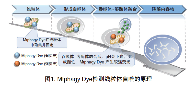
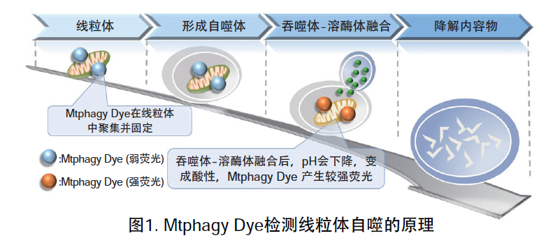
线粒体 (Mitochondria) 是细胞中重要的细胞器之一,可以为细胞活力提供能量 。近年有报道去极化线粒体的积累引起的阿尔茨海默病 (Alzheimer’s Disease) 与帕金森病(Parkinson’s Disease),可能与线粒体自噬有关。线粒体自噬是一种清除机制,可以通过自噬,将氧化应激、DNA损伤因素导致功能失调的线粒体隔离包裹成自噬体(Autophagosome),再与溶酶体 (Lysosome) 融合后降解。本试剂盒内含Mtphagy Dye (用于检测线粒体自噬) 和Lyso Dye (溶酶体染料)。Mtphagy Dye通过化学结合,固定在细胞内的线粒体上,会发出较弱的荧光。当线粒体发生自噬,损伤的线粒体会与溶酶体融合,pH会下降,变成酸性,此时Mtphagy Dye会产生较强的荧光。如想直观观察Mtphagy Dye标记的线粒体和溶酶体的结合,可联合应用试剂盒中的Lyso Dye (标记溶酶体) 进行双染。

特点:

1)只需添加小分子量荧光试剂即可轻松检测线粒体

2)可以使用荧光显微镜进行活细胞成像

3)可以与附着的溶酶体染色剂同时染色

原理

记载了本产品的检测原理和实验例的论文请看MD01论文实验例中第四篇:Live Cell Imaging of Mitochondrial Autophagy with a Novel Fluorescent Small Molecule

线粒体自噬—Mitophagy Detection Kit货号:MD01

实验例

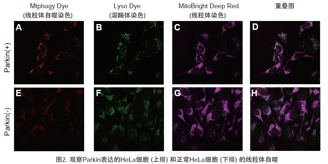
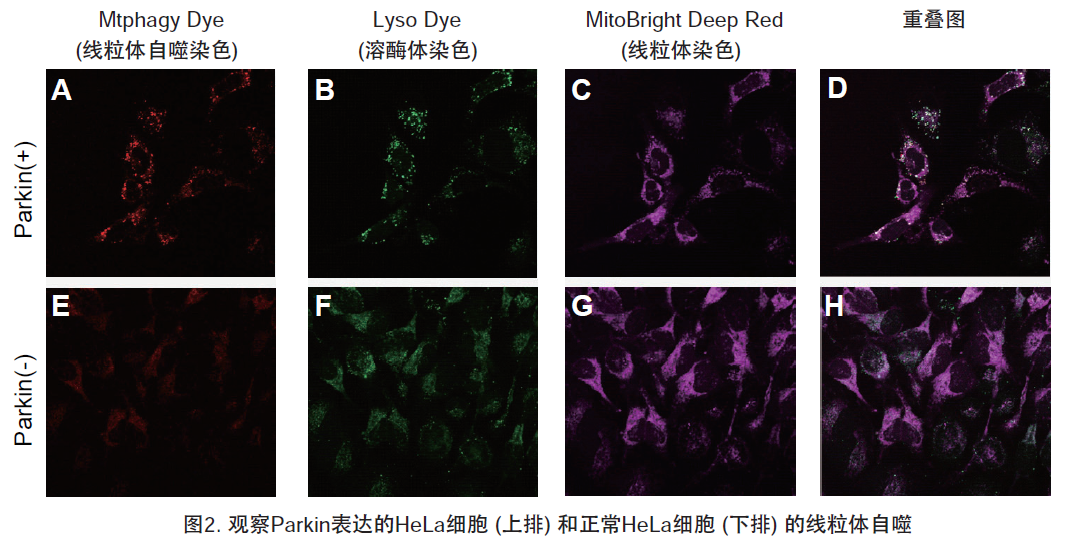
1.用羰基氰化物间氯苯腙 (CCCP,一种线粒体解偶联剂) 诱导Parkin表达的HeLa细胞线粒体自噬,并通过荧光显微镜进行检测。另外,通过与线粒体染色试剂(MitoBright Deep Red:MT08)一同染色,能够区分出已发生自噬的的线粒体(白色)和未发生自噬的线粒体(紫色)(照片:右侧)。

线粒体自噬—Mitophagy Detection Kit货号:MD01

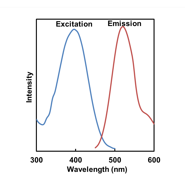
波长:

Mtphagy Dye:561 nm (Ex)、650 LP nm (Em)

Lyso Dye:488 nm (Ex)、502-554 nm (Em)

MitoBright Deep Red:640 nm (Ex)、656-700 nm (Em)

2.荧光显微镜观察

HeLa细胞用CCCP处理,并与线粒体检测试剂(Mtphagy Dye)和线粒体染色试剂(MitoBright LT Green)共同染色,并经过一段时间(6小时)后进行检测。


<检测条件>

设备:LSM-700 Laser scanning confocal microscope (LSCM)

(Carl Zeiss, Oberkochen, Germany)

激发波长:

MitoBright LT Green 488 nm

Mtphagy Dye      555 nm

物镜:63x

拍摄时间:6小时

拍摄间隔:15秒

3.自噬诱导和线粒体膜电位变化关系的检测

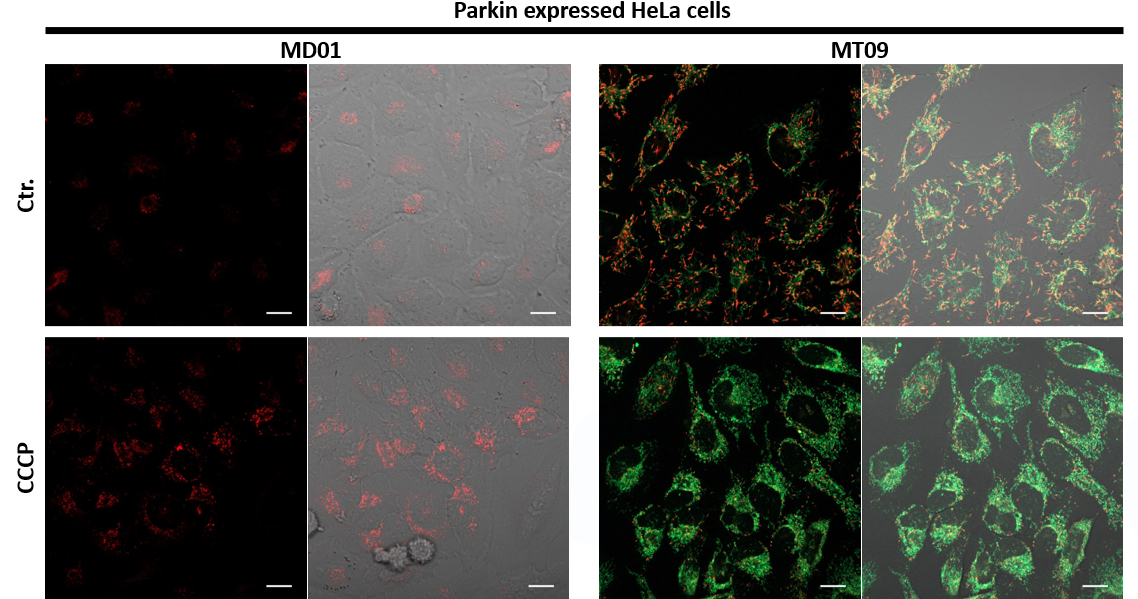
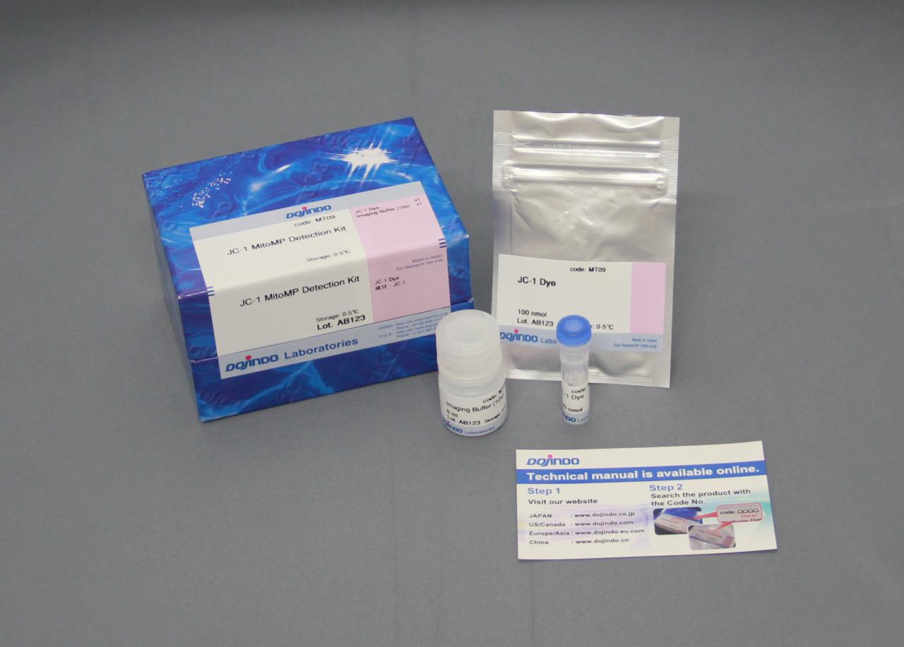
用羰基氰化物间氯苯腙(CCCP,一种线粒体解偶联剂)诱导Parkin表达的HeLa细胞线粒体自噬,并使用线粒体自噬检测试剂盒(Mitophagy Detection Kit:MD01)和线粒体膜电位检测试剂盒(JC-1 MitoMP Detection Kit:MT09)观察荧光结果。

结果证实在未经CCCP处理的细胞中几乎未检测到线粒体自噬的发生,并且线粒体膜电位正常维持。 另一方面,在添加了CCCP的细胞中,证实了线粒体膜电位的降低(JC-1的红色荧光降低)和线粒体自噬的发生(Mtphagy染料的荧光增强)。

<实验条件>

■将Parkin质粒导入HeLa细胞

使用HilyMax(货号:H357)将Parkin质粒引入HeLa细胞中(Parkin质粒/HilyMax试剂:0.1 μg/0.2 μl)

过夜培养后进行检测。

■线粒体自噬检测

向表达Parkin的HeLa细胞中添加0.1 μmol/l Mtphagy工作溶液,并在37°C下孵育30分钟。然后将细胞用HBSS洗涤,加入10 μg/ml CCCP/MEM溶液,并在37℃下孵育2小时。在荧光显微镜下观察细胞。

■线粒体膜电位检测

将10 μg/ml的CCCP/MEM溶液添加至表达Parkin的HeLa细胞中,并在37℃下孵育1.5小时。加入4 μmol/l的JC-1工作液使其终浓度达到2 μmol/l,并在37℃下孵育30分钟。孵育后,将细胞用HBSS洗涤,加入成像缓冲液,并在荧光显微镜下观察细胞。

线粒体自噬—Mitophagy Detection Kit货号:MD01

<检测条件>

■线粒体自噬检测

Ex:561 nm,Em:570-700 nm

■线粒体膜电位检测

绿色Ex:488 nm,Em:500-550 nm

红色Ex:561 nm,Em:560-610 nm

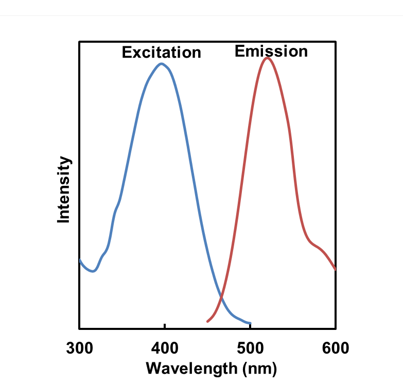
荧光特性

线粒体自噬—Mitophagy Detection Kit货号:MD01

参考文献

序号 检测对象 使用仪器 文献
1) 细胞(HeLa) 流式细胞仪 J. Koniga,   C. Otta, M. Hugoa, T. Junga, A. L. Bulteaub, T. Grunea and A. Hohna, “Mitochondrial contribution to lipofuscin   formation”, Redox Biology, 2017, 11, 673.
2) 细胞(KB) 荧光显微镜 K. Kameyama, “Induction of mitophagy-mediated antitumor activity with   folate-appended methyl-β-cyclodextrin”, International Journal of   Nanomedicine, 2017, 12, 3433.
3) 细胞(SH-SY5Y, 初代皮质神经细胞) 荧光显微镜 E. F. Fang, T. B. Waltz, H.   Kassahun, Q. Lu, J. S. Kerr, M. Morevati, E. M. Fivenson, B. N. Wollman, K.   Marosi, M. A. Wilson, W. B. Iser, D. M. Eckley, Y. Zhang, E. Lehrmann, I. G.   Goldberg, M. S. Knudsen, M. P. Mattson, H. Nilsen, V. A. Bohr and K. G. Becker, “Tomatidine enhances lifespan and healthspan in C. elegans   through mitophagy induction via the SKN-1/Nrf2 pathway”, Scientific   Reports, 2017, 7, (46208), DOI: 10.1038/srep46208.
4) 细胞(HeLa、Parkin表达HeLa) 荧光显微镜 H. Iwashita, S. Torii, N.   Nagahora, M. Ishiyama, K. Shioji, K. Sasamoto, S. Shimizu and K. Okuma, “Live Cell Imaging of Mitochondrial Autophagy with a Novel   Fluorescent Small Molecule”, ACS Chem. Biol., 2017, 12,   (10), 2546.
5) 细胞(Cardiomyocytes) 流式细胞仪 Y. Feng, NB.   Madungwe, CV. da Cruz Junho and JC. Bopassa, “Activation of G protein-coupled oestrogen receptor 1 at   the onset of reperfusion protects the myocardium against ischemia/reperfusion   injury by reducing mitochondrial dysfunction and mitophagy.”, Br.   J. Pharmacol., 2017, 174, (23), 4329.
6) 细胞(HCT116) 荧光显微镜 K. M.   Elamin, K. Motoyama, T. Higashi, Y. Yamashita, A. Tokuda and H. Arima, “Dual targeting system by supramolecular complex of   folate-conjugated methyl-β-cyclodextrin with adamantane-grafted hyaluronic   acid for the treatment of colorectal cancer.”, Int. J. Biol.   Macromol., 2018, doi: 10.1016/j.ijbiomac.2018.02.149.
7) 细胞(Parkin-HeLa) 流式细胞仪 N. Furuya, S. Kakuta, K. Sumiyoshi, M. Ando, R. Nonaka, A. Suzuki, S. Kazuno, S. Saiki   and N. Hattori, “NDP52 interacts with   mitochondrial RNA poly(A) polymerase to promote mitophagy.”, EMBO   Rep. ., 2018, doi: 10.15252/embr.201846363.
8) 细胞(NKT) 流式细胞仪 L. Zhu, X. Xie, L.   Zhang, H. Wang, Z. Jie, X. Zhou, J. Shi, S. Zhao, B. Zhang, X. Cheng and   S. Sun, “TBK-binding protein 1 regulates   IL-15-induced autophagy and NKT cell survival”, Nature   Communications., 2018, 9, (1), doi:10.1038/s41467-018-05097-5.
9) 细胞(HeLa) 流式细胞仪 K. Araki,   K. Kawauchi, W. Sugimoto, D. Tsuda, H. Oda, R. Yoshida and K. Ohtani, “Mitochondrial protein E2F3d, a distinctive E2F3 product,   mediates hypoxia-induced mitophagy in cancer cells”, Commun   Biol., 2019, DOI: 10.1038/s42003-018-0246-9.
10) 细胞(Bovine Sertoli) 荧光显微镜 E. Adegoke, S.   Adeniran, Y. Zeng, X. Wang, H. Wang, C. Wang, H.   Zhang, P. Zheng and G. Zhang , “Pharmacological   inhibition of TLR4/NF-κB with TLR4-IN-C34 attenuated microcystin-leucine   arginine toxicity in bovine Sertoli cells.”, J Appl   Toxicol., 2019,doi: 10.1002/jat.3771.
11) 组织(小鼠) 荧光显微镜  E. F. Fang, Y.   Hou, K. Palikaras, B. A. Adriaanse, J. S. Kerr, B.   Yang, S. Lautrup, M. M. Hasan-Olive, D. Caponio, X.   Dan, P. Rocktaschel, D. L. Croteau, M. Akbari, N. H.   Greig, T. Fladby, H. Nilsen, M. Z. Cader, M. P.   Mattson, N. Tavernarakis and V. A. Bohr, “Mitophagy   inhibits amyloid-β and tau pathology and reverses cognitive deficits in   models of Alzheimer’s   disease.”, Nat. Neurosci. ., 2019,DOI:10.1038/s41593-018-0332-9.
12) 细胞(HepG2) 荧光显微镜 Iwasawa, T.   Shinomiya, N. Ota, N. Shibata, K. Nakata, I. Shiina,   and Y. Nagahara , “Novel Ridaifen-B   Structure Analog Induces Apoptosis and Autophagy Depending on Pyrrolidine   Side Chain”, Biological and Pharmaceutical   Bulletin., 2019, 42, (3), 401-410, doi: 10.1248/bpb.b18-00643.
13) 细胞(U2OS) 荧光显微镜 T. Namba, “BAP31 regulates mitochondrial function via interaction   with Tom40 within ER-mitochondria contact sites “, Sci   Adv., 2019, 5, (6), 1386.
14) 细胞(INS-1) 荧光显微镜 A.   Inamura, S. M. Hirayama, and K. Sakurai, Loss of   Mitochondrial DNA by Gemcitabine Triggers Mitophagy and Cell   Death’, Biol. Pharm. Bull.., 2019, 42, 1977.
15) 细胞(HRCEpiC, HRPTEpic) 流式细胞仪 Y. Zhao and   M. Sun, Metformin rescues Parkin protein expression   and mitophagy in high glucose-challenged human renal epithelial cells by   inhibiting NF-κB via PP2A activation., Life   Sci.., 2020, DOI:10.1016/j.lfs.2020.117382.
16) 细胞(RAES) 荧光显微镜 N. Liu, J. Wu, L. Zhang, Z. Gao,   Y. Sun, M. Yu, Y. Zhao, S. Dong, F. Lu and W. Zhang , “Hydrogen Sulphide modulating mitochondrial morphology to   promote mitophagy in endothelial cells under high‐glucose and high‐palmitate   “, J. Cell. Mol. Med., 2017, 21, (12), 3190.
17) 细胞(BAECs) 荧光显微镜 N. Kajihara, D. Kukidome, K.   Sada, H. Motoshima, N. Furukawa, T. Matsumura, T. Nishikawa and E.   Araki, “Low glucose induces mitochondrial   reactive oxygen species via fatty acid oxidation in bovine aortic endothelial   cells”, J Diabetes Investig, 2017, 8, (6), 750.
18) 细胞(HT22) 荧光显微镜 M. Jin, H. Ni and  L.   Li, “Leptin Maintained Zinc Homeostasis Against   Glutamate-Induced Excitotoxicity by Preventing Mitophagy-Mediated   Mitochondrial Activation in HT22 Hippocampal Neuronal   Cells.”, Front Neurol, 2018, 9, (9), 332.
19) 细胞(BMDMs) 流式细胞仪 D. Bhatia, K. P. Chung, K.   Nakahira, E. Patino, M. C. Rice, L. K. Torres, T. Muthukumar, A. M. Choi, O.   M. Akchurin and M. E. Choi , “Mitophagy-dependent   macrophage reprogramming protects against kidney fibrosis”, JCI   Insight, 2019, 4, (23), e132826.
20) 细胞(U2OS) 荧光显微镜 J. Zheng, D. L. Croteau, V. A.    Bohr and M. Akbari, “Diminished OPA1   expression and impaired mitochondrial morphology and homeostasis in   Aprataxin-deficient cells. “, Nucleic Acids   Res., 2019, 47, (8), 4086.
21) 细胞(HT22) 荧光显微镜 D. D. Wang, M. F. Jin, D. J. Zhao   and H. Ni, “Reduction of Mitophagy-Related   Oxidative Stress and Preservation of Mitochondria Function Using Melatonin   Therapy in an HT22 Hippocampal Neuronal Cell Model of Glutamate-Induced   Excitotoxicity”, Front Endocrinol (Lausanne), 2019, 10,   550.
22) 细胞(CD4+T-cells, HeLa) 荧光显微镜 A. Bektas, S. H. Schurman, M. G.   Freire, A. Bektas, S. H. Schurman, M. G. Freire, C. A. Dunn, A. K. Singh, F.   Macian, A. M. Cuervo, R. Sen and L. Ferrucci, “Age-associated   changes in human CD4+ T cells point to mitochondrial dysfunction consequent   to impaired autophagy.”, Aging (Albany NY)., 2019, 11,   (21), 9234-9263.
23) 细胞(ALM) 流式细胞仪 T. Nechiporuk, S.E. Kurtz, O.   Nikolova, T. Liu, C.L. Jones, A. D. Alessandro, R. C. Hill, A. Almeida, S. K.   Joshi, M. Rosenberg, C. E. Tognon, A. V. Danilov, B. J. Druker, B. H. Chang,   S. K McWeeney and J. W. Tyner, “The TP53   Apoptotic Network Is a Primary Mediator of Resistance to BCL2 Inhibition in   AML Cells.”, Cancer Discov., 2019, 9, (7), 919.
24) 细胞(PK-15) 荧光显微镜 Y. Zhang, R. Sun, X. Li  and   W. Fang, “Porcine Circovirus 2 Induction of ROS Is Responsible for Mitophagy in PK-15 Cells via Activation of Drp1 Phosphorylation”, Viruses., 2020, 12, (3), 289.
25) 细胞(HCE) 荧光显微镜 Y. Huo, W. Chen, X. Zheng, J.   Zhao, Q. Zhang, Y. Hou, Y. Cai, X. Lu and X. Jin , “The protective effect of EGF-activated ROS in human   corneal epithelial cells by inducing mitochondrial autophagy via activation   TRPM2.”, J. Cell. Physiol., 2020, DOI: 10.1002/jcp.29597.
26) 细胞(心肌细胞) 荧光显微镜 Y. Sun, F. Lu, X. Yu, B. Wang, J.   Chen, F. Lu, S. Peng, X. Sun, M. Yu, H. Chen, Y. Wang, L. Zhang, N. Liu, H.   Du, D. Zhao and W. Zhang, “Exogenous H2S   Promoted USP8 Sulfhydration to Regulate Mitophagy in the Hearts of db/db   Mice.”, Aging Dis., 2020, 11, (2), 269.
27) 细胞(HCFs) 荧光显微镜 R. Tanaka, M. Umemura, M.   Narikawa, M. Hikichi, K. Osaw, T. Fujita, U. Yokoyama, T. Ishigami, K. Tamura   and Y. Ishikawa, “Reactive fibrosis precedes   doxorubicin-induced heart failure through sterile   inflammation.”, ESC Heart Fail., 2020, 7, (2), 588.
28) 细胞(VSMCs) 荧光显微镜 C. Duan, L. Kuang, X. Xiang, J.   Zhang, Y. Zhu, Y. Wu, Q. Yan, L. Liu and T. Li, “Drp1   regulates mitochondrial dysfunction and dysregulated metabolism in ischemic   injury via Clec16a-, BAX-, and GSH- pathways “, Cell Death   Dis., 2020, 11, 251.
29) 细胞(Bovine Sertoli) 荧光显微镜 E. O. Adegoke, W. Xue, N. S.   Machebe, S. O. Adeniran, W. Hao, W. Chen, Z. Han, Z. Guixue and Z.   Peng, “Sodium Selenite inhibits mitophagy,   downregulation and mislocalization of blood-testis barrier proteins of bovine   Sertoli cell exposed to microcystin-leucine arginine (MC-LR) via TLR4/NF-kB   and mitochondrial signaling pathways blockage.”, Ecotoxicol.   Environ. Saf., 2018, 116, 165.
30) 细胞(HeLa) 荧光显微镜 D. Takahashi, J. Moriyama, T.   Nakamura, E. Miki, E. Takahashi, A. Sato, T. Akaike, K. I. Nakama and H.   Arimoto, “AUTACs: Cargo-Specific Degraders   Using Selective Autophagy. “, Mol. Cell, 2019, 76,   (5), 797.
31) 细胞(primary hepatocyte) 荧光显微镜 H. Kim, J. H. Lee and J. W.   Park, “IDH2 deficiency exacerbates   acetaminophen hepatotoxicity in mice via mitochondrial dysfunction-induced   apoptosis.”, Biochim Biophys Acta Mol Basis   Dis, 2019, 1865, (9), 2333.
32) 细胞(C3H10T1/2s) 荧光显微镜 M. S.    Rahman and Y. S.  Kim, “PINK1-PRKN   mitophagy suppression by Mangiferin promotes a brown-fat-phenotype via   PKA-p38 MAPK signalling in murine   C3H10T1/2”, Metabolism, 2020, 101, 154228.
33) 细胞(NHEKs) 荧光显微镜 S. Ikeoka   and A. Kiso  , “The Involvement of   Mitophagy in the Prevention of UV-B-Induced Damage in Human Epidermal   Keratinocytes “, J. Soc. Cosmet. Chem.   Jpn., 2020,  54(3), 252.

常见问题Q&A


Q1: 本试剂盒和现存传统方法相比有何优势?

A1: 与PH敏感并基于Keima荧光蛋白检测方法相比,本试剂为小分子荧光试剂,因此无需表达荧光蛋白。

另外,可以通过与用于普通活细胞成像的荧光试剂用相同的操作方法对其进行染色和共同观察。

Q2: 使用DMSO配置后的储存液稳定性如何?
A2:Mtphagy Dye、Lyso Dye均在制备后需保存在-20℃情况下可以稳定保存1个月。建议按照实验用量,

提前分装保存。

Q3: 工作液稳定性如何
A3: 无法保存,建议现配现用。
Q4: 培养基中有酚红会影响检测吗?
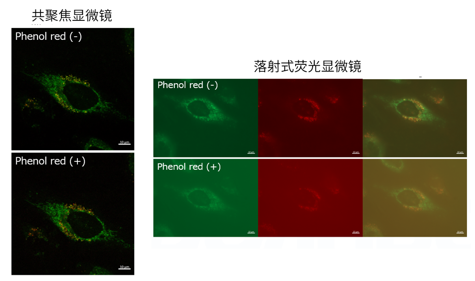
A4:观察的时候,如果使用共聚焦激光显微镜的话,几乎不会受到酚红的影响,但是使用落射型荧光显微

镜的话,会观察到酚红色的背景。(参照以下观察数据)因此使用落射型荧光显微镜时,请在Working

solution进行染色时使用不含酚红的培养基或HBSS。

线粒体自噬—Mitophagy Detection Kit货号:MD01

Q5: 荧光显微镜推荐的滤镜是什么?
A5:根据各种试剂推荐以下波长。Mtphagy Dye:激发(500~560 nm)、发射(670~730 nm)

Lyso Dye:激发(350~450 nm)、发射(500~560 nm)

Q6:与其他深红色染料共同染色时的注意事项。
A6:Mtphagy Dye比一般的红色系荧光染料相比波长更长,所以和Deep Red的荧光染料一起染色的时候

需要特别注意。即Mtphagy Dye在500–560 nm处激发,可在670-730 nm处检测到荧光,这时与

MitoBright  Deep Red的荧光检测波长重叠。因此,有必要在不激发深红色染料的波长下激发Mtphagy

染料,同时在不激发Mtphagy染料的波长下激发深红色染料。

[泄漏的情况]

① 制备仅添加了MitoBright Deep Red(没有添加Mtphagy Dye)的细胞。

② 通过观察MitoBright Deep Red的激发/发射波长,确认是否观察到荧光(右下图)。

③ 用Mtphagy Dye的激发/发射波长观察,确认是否观察到荧光(左下图)。

和③中,观察到来自MitoBright Deep Red的荧光(左下图)。

*如果如上所述确认荧光泄漏,请参阅以下内容。

○调整激发/发射波长

如以上确认如图所示,MitoBright Deep Red也在Ex 561 nm处激发,因此可以将Mtphagy Dye的激发

波长更改为接近激光器或滤光片的500 nm,以使MitoBright深红色不被激发。

调整荧光强度和荧光检测灵敏度

如果MitoBright Deep Red的荧光泄漏到Mtphagy染料的观察波长中,请将观察过程中的激发强度或

灵敏度降低到未观察到荧光的水平。

然后,再确认改变后的观察条件下可以检测Mtphagy Dye的荧光。

[如何检查泄漏]

使用Mtphagy Dye,Lyso dye(溶酶体染色剂),MitoBrightLT Deep Red(线粒体染色剂)

进行三重染色时进行确认

1.在3个培养皿或孔中制备细胞。

(Mtphagy Dye、Lyso Dye、MitoBright Deep Red分别在在不同的皿或孔中进行染色)

2.向每个孔中添加Mtphagy Dye和MitoBright Deep Red。 (在无血清培养基中)

3.在37°C下孵育30分钟。

4.进行自噬诱导条件下(如饥饿培养等)进行培养。

5.向上述2.中未使用的细胞添加Lyso Dye。(在无血清培养基中)

6.在37°C下孵育30分钟。

7.观察每种试剂的激发波长和荧光波长以及荧光强度。

8.检查所用试剂以外的观察波长处的荧光是否没有泄漏。

[观察条件]

Lyso Dye:Ex:350-450 nm,Em:500-560 nm

Mtphagy Dye:Ex : 500-560 nm,Em :670-730 nm

MitoBright Deep Red:Ex :640 nm,Em :656-700 nm

关联产品

线粒体自噬—Mitophagy Detection Kit货号:MD01
线粒体膜电位检测试剂盒—JC-1 MitoMP Detection Kit
线粒体膜电位检测试剂盒

线粒体自噬—Mitophagy Detection Kit货号:MD01
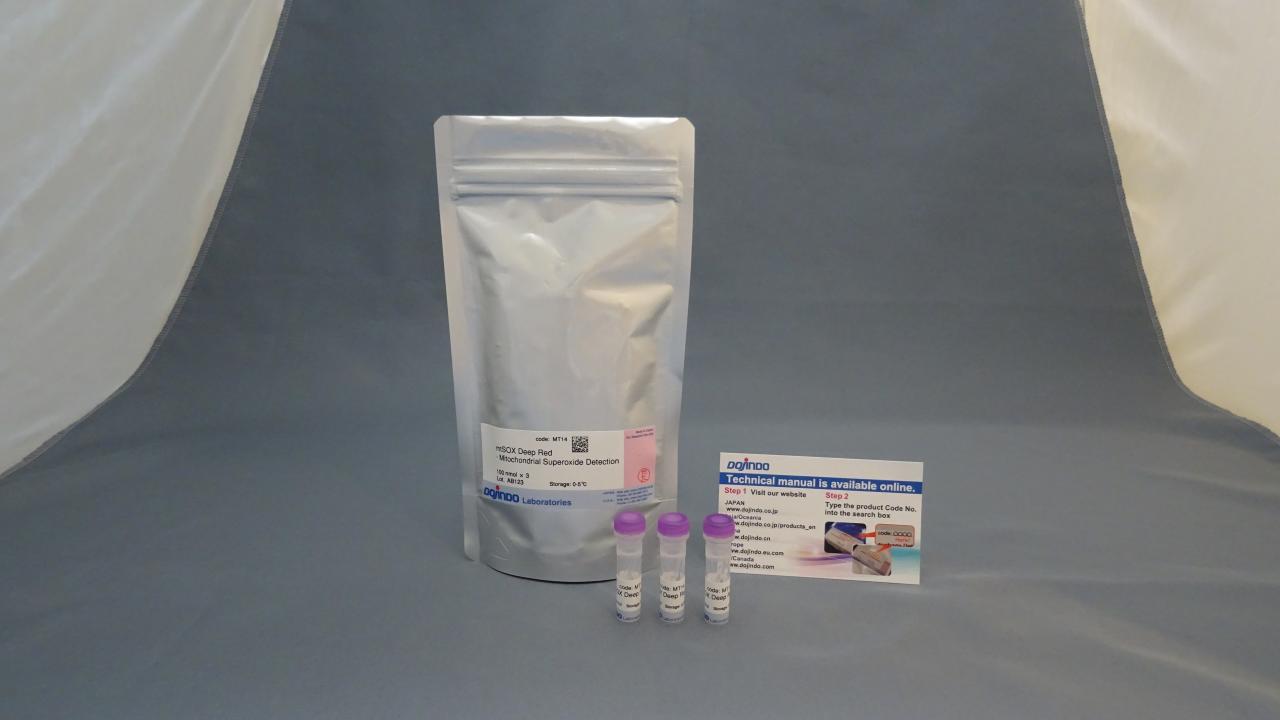
mtSOX Deep Red – Mitochondrial Superoxide Detection
线粒体超氧化物检测用荧光染料

线粒体自噬—Mitophagy Detection Kit货号:MD01
线粒体膜电位检测试剂盒
线粒体膜电位检测试剂盒

线粒体自噬—Mitophagy Detection Kit货号:MD01
MitoBright LT Green试剂
线粒体长效荧光探针-绿色

线粒体自噬—Mitophagy Detection Kit货号:MD01
MitoBright LT Deep Red试剂
线粒体长效荧光探针-深红色

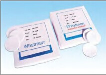
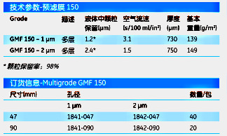
Whatman1842-047Multigrade GMF 150 预滤膜MULTIGRADE 2um 4.7CM 40/PK

【简单介绍】

Whatman GMF150是一种多层玻璃微纤维滤膜,最上层是

个粗滤层(10µm),下层是孔径为1µm或者2µm的有网孔的细

滤层。采用100%硼硅酸玻璃纤维制成,没有任何添加剂,

通过很好的预滤作用来增强对颗粒的负载能力和加快过滤

速度。

【简单介绍】

Whatman GMF150是一种多层玻璃微纤维滤膜,最上层是

个粗滤层(10µm),下层是孔径为1µm或者2µm的有网孔的细

滤层。采用100%硼硅酸玻璃纤维制成,没有任何添加剂,

通过很好的预滤作用来增强对颗粒的负载能力和加快过滤

速度。

【详细说明】

原装进口英国Whatman1842-047Multigrade GMF 150 预滤膜MULTIGRADE 2um 4.7CM 40/PK

英国Whatman1842-047Multigrade GMF 150 预滤膜MULTIGRADE 2um 4.7CM 40/PK

产品介绍:

Multigrade GMF 150预滤膜

Whatman GMF150是一种多层玻璃微纤维滤膜,最上层是

个粗滤层(10μm),下层是孔径为1μm或者2μm的有网孔的细

滤层。采用100%硼硅酸玻璃纤维制成,没有任何添加剂,

通过很好的预滤作用来增强对颗粒的负载能力和加快过滤

速度。

GMF 150 允许:

l 更高的负载力。

l 更快的流速。

l 延长滤器的寿命。

多样孔结构,更好的过滤效率

GMF 150代表了分离实验一个新的领域,最重要的是更快

和更经济的过滤。在应用中,GMF在孔内或者粗滤层表面截

留较大颗粒,而中等大小颗粒则停留在孔状层的表面,最

小的颗粒则滤在微滤层的空隙里。

英国Whatman1842-047Multigrade GMF 150 预滤膜MULTIGRADE 2um 4.7CM 40/PK

上海金畔生物科技有限公司

文章号19708643-19708643

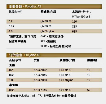
Whatman6724-5145Polydisc AS在线滤器POLYDISC AS 0.45 GF/PES 50/PK

【简单介绍】

Whatman为客户提供多种多样的特殊实验产品,以满足实验者的不同实验需求。秉承Whatman传统的质量,这些产品集合了易于操作、精确度高、一致性好等优点。

【简单介绍】

Whatman为客户提供多种多样的特殊实验产品,以满足实验者的不同实验需求。秉承Whatman传统的质量,这些产品集合了易于操作、精确度高、一致性好等优点。

【详细说明】

原装进口英国Whatman6724-5145Polydisc AS在线滤器POLYDISC AS 0.45 GF/PES 50/PK

英国Whatman6724-5145Polydisc AS在线滤器POLYDISC AS 0.45 GF/PES 50/PK

产品介绍:

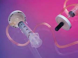
Clyde在线滤器

过滤除菌和分装组织培养基最方便的方法。

l 0.2μm孔径,可连续过滤除菌2 L样品

l 完全一体化

l 一体化的针头泵提供正压,可以在任何场所进行过滤

l 包括软管和单向检测阀

l 玻璃微纤维预滤器

l 射线灭菌独立包装

l 不产生高热,保证生物安全

Clyde™内为不对称混合纤维素酯膜,过滤区域16 cm2,外壳

是丙烯酸材料,其他部分材质为:聚丙烯、PVC和聚乙烯。过

滤量20 ml

英国Whatman6724-5145Polydisc AS在线滤器POLYDISC AS 0.45 GF/PES 50/PK

英国Whatman6724-5145Polydisc AS在线滤器POLYDISC AS 0.45 GF/PES 50/PK
Clyde可以过滤培养基以及和纤维素膜兼容的水相溶液。使

Clyde不需真空泵或其他外力,这使户外操作成为可能。

在线滤器

Whatman在线过滤的特点是具有高纯度的聚丙稀外壳,保证

了样品的纯度,并且针对不同的水溶液和有机溶剂,有多种

过滤介质可供选择。这些滤器使用最先进的构造方法和设计

特点,这种水平的设计提供了最好的一次性再行过滤装置。

这种装置可以高压灭菌,并且有灭菌包装可供选择。

Whatman在线过滤/脱气装置(IFD)直接连入HPLC管线,可

以对流动相同时进行脱气和过滤。

Polydisc滤器

Whatman Polydisc50 mm在线盘状滤器专为航天行业或生

产行业中实验室大容量样品过滤而设计。这种滤器用途广泛,

而且经济实惠。1个装置的过滤量可高达1升。Polydisc滤器可

与注射器或通过阶梯状长管连接的在线装置联合使用。

Polydisc滤器由高纯度的聚丙稀外壳制成,保证了样品的纯

度,而且有各种过滤介质供您选择来适合不同的水样品和有

机样品。

Polydisc AS

Polydisc AS(水溶液)50 mm滤器系列有高通量的聚醚砜膜,蛋

白吸附性低,没有表面活性剂,可用于医药工业。玻璃微纤

维预滤层延长了膜的寿命,并有效过滤高污染样品。每一个

Polydisc AS滤器在出口处都有一个灭菌的罩子。用“医用级”

洁净灭菌包装密封,确保使用安全。

特点

l 放射灭菌,无EtO残留

l 倒钩状接口,适合多种管径

l 完整性测试(气泡点)

l 重量很轻(11.5 g):避免了因滤器过重而引起的软管塌陷

应用

l 组织培养介质

l 试剂制备

l 颗粒计数溶液

l 药物准备

英国Whatman6724-5145Polydisc AS在线滤器POLYDISC AS 0.45 GF/PES 50/PK

上海金畔生物科技有限公司

文章号19837167-19837167

上海金畔生物科技有限公司提供Axygen常规耗清单T-200-Y200ul黄色吸头

上海金畔生物科技有限公司提供Axygen常规耗清单T-200-Y200ul黄色吸头

货号 产品名称 销售包装 销售单位 目录价(销售包装)
T-300 10ul透明吸头 1000 Tips / 包, 20 包 / 箱 157.2
T-200-Y 200ul黄色吸头 1000 Tips / 包, 20 包 / 箱 150.6
T-1000-B 1000ul蓝色吸头 1000 Tips / 包, 5 包 / 箱 162.6
MCT-150-C 1.5ml无色离心管 500 Tubes / 包, 10 包 / 箱 122.7
PCR-02-C 0.2ml透明平盖PCR薄壁管 1000 Tubes / 包, 10 包 / 箱 453.8
PCR-0208-C 0.2ml 八连排透明PCR薄壁管 125 Strips / 包, 10 包 / 箱 907.6
PCR-2CP-RT-C 0.2ml透明PCR八排管平盖(荧光定量专用) 125 Strips / 包, 10 包 / 箱 352.9


T-200-C 200ul 透明吸头 1000 Tips / 包, 20 包 / 箱 160.7
T-200-C-R 200ul 盒装透明吸头 10 盒 / 包, 5 包 / 箱 36.3
T-200-C-R-S 200ul 盒装灭菌透明吸头 10 盒 / 包, 5 包 / 箱 41.9
T-200-C-STK 200ul 叠装透明吸头 5 小叠 / 叠, 10叠 / 箱 181.7
T-200-C-STK-S 200ul 叠装灭菌透明吸头 5 小叠 / 盒, 10 盒 / 箱 209.6
T-200-Y 200ul黄色吸头 1000 Tips / 包, 20 包 / 箱 150.6
T-200-Y-R 200ul 盒装黄吸头 10 盒 / 包, 5 包 / 箱 36.3
T-200-Y-R-S 200ul 盒装灭菌黄吸头 10 盒 / 包, 5 包 / 箱 41.6
T-200-Y-STK 200ul叠装黄色吸头 5 小叠 / 叠, 10叠 / 箱 181.7
T-200-Y-STK-S 200ul叠装灭菌黄色吸头 5 小叠 / 叠, 10叠 / 箱 209.6
T-205-WB-C 200ul宽嘴透明吸头 1000 Tips / 包, 20 包 / 箱 160.7
T-205-WB-C-R 200ul宽嘴盒装透明吸头 10 盒 / 包, 5 包 / 箱 39.1
T-205-WB-C-R-S 200ul宽嘴盒装灭菌透明吸头 10 盒 / 包, 5 包 / 箱 44.7
T-210-C 200ul透明平嘴吸头 1000 Tips / 包, 20 包 / 箱 160.7
T-210-C-R 200ul盒装透明平嘴吸头 10 盒 / 包, 5 包 / 箱 36.3
T-210-C-R-S 200ul盒装灭菌透明平嘴吸头 10 盒 / 包, 5 包 / 箱 41.9
T-210-Y 200ul黄色平嘴吸头 1000 Tips / 包, 20 包 / 箱 160.7
T-210-Y-R 200ul 盒装黄色平嘴吸头 10 盒 / 包, 5 包 / 箱 36.3
T-210-Y-R-S 200ul 盒装灭菌黄色平嘴吸头 10 盒 / 包, 5 包 / 箱 41.9
T-300 10ul透明吸头 1000 Tips / 包, 20 包 / 箱 157.2
T-300-R 0.5-10ul盒装透明Gilson吸头 10 盒 / 包, 5 包 / 箱 36.3
T-300-R-S 0.5-10ul盒装灭菌透明Gilson吸头 10 盒 / 包, 5 包 / 箱 41.9
T-300-STK 0.5-10ul叠装透明Gilson吸头 5 小叠 / 叠, 10叠 / 箱 181.7
T-300-STK-S 0.5-10ul叠装灭菌透明Gilson吸头 5 小叠 / 叠, 10叠 / 箱 218.0
T-350-C 300ul透明吸头 1000 Tips / 包, 10 包 / 箱 195.6
T-350-C-R 300ul 盒装透明吸头 10 盒 / 包, 5 包 / 箱 41.9
T-350-C-R-S 300ul 盒装灭菌透明吸头 10 盒 / 包, 5 包 / 箱 47.5
T-384-R 50ul盒装透明吸头 10 盒 / 包, 5 包 / 箱 726.6
T-384-R-S 50ul盒装灭菌透明吸头 10 盒 / 包, 5 包 / 箱 810.5
T-400 0.5-10ul透明Eppendorf吸头 1000 Tips / 包, 20 包 / 箱 167.7
T-400-R 0.5-10ul盒装透明Eppendorf吸头 10 盒 / 包, 5 包 / 箱 36.3
T-400-R-S 0.5-10ul盒装灭菌透明Eppendorf吸头 10 盒 / 包, 5 包 / 箱 41.9
T-5000-C 1000-5000ul透明吸头 250 Tips / bag, 10 bags / 箱 263.0

 
更多产品,更多优惠,请联系我们!
上海金畔生物科技有限公司
地 址: 上海市浦东新区东靖路699弄36栋701室
邮 编: 201208
固话总机:
订货热线:15221999938
网 址: www.jinpanbio.com
金畔博客:www.jinpanbio.cn
Email:sales@jinpanbio.com