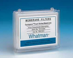
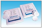
Whatman112104Nuclepore径迹蚀刻膜 NUC PC 142MM 0.08um 25/PK

【简单介绍】

Whatman给实验者提供了多种材质的滤膜,先进的技术规范使得今天很多的应用都更愿意选择Whatman
产品。滤膜的孔径精确、韧性好,保证了再现性和一致性。Whatman滤膜孔径范围很广,从0.2µm-12µm,并有很多选择的膜过滤容器。
无菌和消毒包装可用于特殊应用中,也有着色网格类型可供选择。

【简单介绍】

Whatman给实验者提供了多种材质的滤膜,先进的技术规范使得今天很多的应用都更愿意选择Whatman
产品。滤膜的孔径精确、韧性好,保证了再现性和一致性。Whatman滤膜孔径范围很广,从0.2µm-12µm,并有很多选择的膜过滤容器。
无菌和消毒包装可用于特殊应用中,也有着色网格类型可供选择。

【详细说明】

原装进口英国Whatman112104Nuclepore径迹蚀刻膜 NUC PC 142MM 0.08um 25/PK

英国Whatman112104Nuclepore径迹蚀刻膜 NUC PC 142MM 0.08um 25/PK

产品介绍:

Whatman给实验者提供了多种材质的滤膜,先进的技术规范使得今天很多的应用都更愿意选择Whatman
产品。滤膜的孔径精确、韧性好,保证了再现性和一致性。Whatman滤膜孔径范围很广,从0.2μm-12μm,并有很多选择的膜过滤容器。
无菌和消毒包装可用于特殊应用中,也有着色网格类型可供选择。

产品特点:

Nuclepore聚碳酸径迹蚀刻膜由高质量的聚碳酸薄膜制成,具有孔径精确、流速快等特点,并且有着极好的耐化学性和耐热性,这类膜表面光滑而平整,对提取物几乎没有吸附。这种膜还有黑色膜,背景反差大,用于荧光和其它显微镜法。

Track-Etched Polycarbonate and Polyester Membranes

径迹蚀刻聚碳酸酯膜和聚脂膜

Nuclepore Track-Etched Membranes聚碳酸酯膜

Nuclepore聚碳酸酯径迹蚀刻膜是由高品质的聚碳酸酯薄膜制成,具有孔径精确、流速快等特点。并且有着极好的耐化学性和耐热性。这类膜表面光滑而平整,对提取物几乎没有吸附。

特性

不吸附蛋白和提取物,对样品无污染

良好的耐化学性和热稳定性,适用于大多数样品

低灰分、低皮重

膜表面光滑而平整,便于对截留物粒子进行观察

可提供黑色膜,用于荧光和其他纤维镜法,背景反差大

应用

荧光显微镜、生物分析、环境分析、寄生生物学、细胞培养、空气分析、EPA检验、

水体微生物学、燃烧检测。

技术参数-Nuclepore聚碳酸酯膜

产品名

聚碳酸酯

厚度

6-11μm

爆裂强度

>10 psi

净重

0.6-1mg/ cm2

原料比重

1.20g/ cm2

热封范围

230-275

最高可耐高温

140

易燃性

Slow burn

灰度

0.92μg/ cm2

孔率

<15%

计算孔径

0.05-12.0μm

计算孔密度

10*5-6×10*8pore/ cm2

表面质地

平滑

透明度

半透明

折射率

1.584-1.625(双折射)

疏水

纤维释放

高温蒸汽灭菌

121

订货信息-Nuclepore聚碳酸酯膜

直径(mm

孔径(um

货号

直径(mm

孔径(um

货号

47

0.015

111101

90

0.2

111706**

47

0.05

111103

90

0.4

111707**

47

0.08

111104

90

1

111710**

47

0.1

111105

90

2

111711**

47

0.2

111106

142

0.08

112104**

47

0.4

111107

142

0.1

112105**

47

0.6

111108

142

0.2

112106**

47

0.8

111109

142

0.4

112107**

47

1

111110

142

0.6

112108**

47

2

111111

142

1

112110**

47

3

111112

293

0.2

112806**

47

5

111113

293

0.4

112807**

47

8

111114

293

1

112810**

47

10

111115

293

2

112811**

47

12

111116

8×10

0.03

113502**

47*

0.4

111137

19×42

5

113313

47***

0.4

111130

25×80**

8

155846

*AOX―适用于AOXAdsorbable Organic Halogens)分析

**不含PVP疏水 ****Good Coated PC *50/

***AERO **25/

***10/

上海金畔生物科技有限公司

文章号19716224-19716224

ES/iPS细胞分化诱导研究用低分子化合物

ES/iPS细胞分化诱导研究用低分子化合物

  • 产品特性
  • 相关资料
  • Q&A
  • 参考文献

ES/iPS细胞分化诱导研究用低分子化合物ES/iPS细胞分化诱导研究用低分子化合物


  Wako提供大量ES/iPS未分化维持、分化诱导相关的低分子化合物,推出CultureSure® A419259、三盐酸化物和CKl-7二盐酸化物,MF 、CultureSure® KY03-Ⅰ等新品。


● ES·iPS细胞的分化诱导、脱分化等。


产品编号

品名

规格

容量

作 用

034-24801

CultureSure® A419259 Trihydrochloride

细胞培养用

1mg

030-24803

5mg

038-24804

25mg

034-24806

100mg

Src族抑制剂。Src族激酶在细胞遗传、分化、增殖相关的信号传递起着重要作用。

011-25291

A-769662

细胞生物学用

5mg

017-25293

25mg

AMPK活性化剂。AMPK活性化对促进细胞生长、增殖、生存的mTOR信号传递有抑制作用。

015-22531

AICAR

细胞生物学用

100mg

011-22533

1g

AMPK催化剂。抑制脂肪细胞分化,将神经干细胞诱导为星型胶质细胞。〔Giri, S., et al.: Nutr. Metab. (Lond), (2006).〕〔Zang, Y., et al.: J. Biol. Chem., 283, 6201 (2008). 〕

014-16631

Am580

生化学用

5mg

合成类维生素A。比生长因子能更快速高效地将人iPS细胞和ES细胞诱导为中间中胚层。〔Araoka, T., et al.: PLOS ONE, 9, e84881 (2014).〕

014-25421

AMD3100 Octahydrochloride

细胞生物学用

10mg

CXCR4拮抗剂。可高效调动体内造血干细胞。〔Lura, A., et al., : Exp Hematol., 39(3), 384 (2011).〕

035-16303

Ciclosporin A

生化学用

100mg

039-16301

200mg

提高小鼠/人iPS细胞到心肌细胞和心肌前驱细胞的诱导效率。〔Fujiwara, M., et al.: PLOS ONE, 6, e16734 (2006).〕

030-20981

Ciglitazone

细胞生物学用

5mg

PPARγ激动剂。诱导人类间充质干细胞分化成脂肪细胞、抑制成骨纤维细胞分化。〔Benvenuti, S., et al.: J. Endocrinol. Invest., 30, RC26 (2007).〕

034-21501

CKI-7 Dihydrochloride

细胞生物学用

5mg

蛋白激酶(CK1)抑制剂。在无血清无饲养细胞培养基中与SB431542, Y-27632结合使用,将人ES/iPS细胞分化诱导成视网膜前驱细胞。〔Takahashi, M., et al.: J. Cell Sci., 122, 3169 (2009).〕

043-33581

DAP【γ-Secretase InhibitorⅨ】

  细胞生物学用

5mg

γ-Secretase抑制剂。抑制Notch信号,诱导ES细胞胚状体分化为神经。〔Nelson, BR., et al.: Dev. Biol., 304, 479 (2007).〕〔Crawford, TQ., et al.: Dev. Dyn., 236, 886 (2007).〕

067-02191

Forskolin

毛喉素

生化学用

10mg

063-02193

25mg

腺苷酸环化酶催化剂。与bFGF一起使用,可诱导间充质干细胞分化为神经细胞。〔Jang, S. et   al.: BMC Cell Biol., 11, 25 (2010).〕

034-24301

CultureSure® IWP-2

细胞培养用

5mg

030-24303

25mg

通过Porcupine(Porcn)的失活抑制Wnt蛋白质的棕榈酰化,促进人iPS细胞分化为心肌细胞。已经支原体、细胞毒性检测确认。Minami, I. et al.:   Cell Rep., 2, 1448 (2012).〕

094-06381

IWR-1-endo

细胞生物学用

5mg

Wnt信号抑制剂。稳定分解β- catenin的复合体(由Axin2, Apc, Ck1, Gsk3β组成),促进β- catenin的分解。

124-06011

LDN193189 Hydrochloride

LDN193189盐酸盐

细胞生物学用

2mg

BMP I型受体/ALK2,ALK3抑制剂。调节BMP信号的产生,在成熟组织中起着极其重要的作用。〔Yu, P.B., et al.: Nat. Med., 14, 1363 (2008).〕

129-04861

LY294002

生化学用

5mg

125-04863

10mg

123-04864

25mg

Phosphatidylinositol 3-kinase抑制剂。诱导小鼠ES细胞分化为胰岛细胞(胰岛素产生细胞)。

166-23991

Purmorphamine

2,6,9-三取代嘌呤化合物,PM

细胞生物学用

5mg

Hedgehog信号激动剂。诱导间充质前驱细胞或前造骨性细胞分化成成骨细胞。〔Wu, X., et al.: Chem. Biol., 11, 1229 (2004).〕

186-01114

all -trans-Retinoic Acid

全反式维甲酸

生化学用

50mg

182-01116

100mg

182-01111

250mg

188-01113

1g

诱导神经细胞的分化。

184-02391

Reversine

细胞生物学用

5mg

人A3腺苷受体激动剂。成肌细胞脱分化为干细胞。〔Chen, S., et al.: J. Am. Chem. Soc., 126, 410 (2004).〕

192-16541

SB431542

细胞生物学用

5mg

198-16543

25mg

ALK4, ALK5, ALK7抑制剂。促进ES细胞源内皮细胞的增殖、分化、膜形成。本品和PD0325901、Thiazovivin使用的话,可将细胞重组效率提高200倍以上,加速细胞重组。

〔Lin T., et al.: Nat. Methods, 6, 805 (2009).〕〔Ogawa, K., et al.: J. Cell. Sci.,120, 55 (2007).〕

198-09811

Spermine

精胺

生化学用

250mg

194-09813

1g

诱导心肌细胞分化。〔Sasaki, T., et al.: Differentiation, 76, 1023 (2008).〕

203-17561

Trichostatin A

曲古柳菌素A

细胞生物学用

1mg

209-17563

5mg

组蛋白脱乙酰基酶抑制剂。诱导心肌细胞分化。〔Oyama, T., et al.: J.   Cell. Biol., 176, 329 (2007).〕

209-19481

Troglitazone

曲格列酮

细胞生物学用

5mg

205-19483

50mg

PPARγ激动剂。诱导人间充质干细胞分化为脂肪细胞,抑制其分化为成骨细胞。〔Benvenuti, S., et al.: J. Endocrinol. Invest., 30, RC26 (2007).〕

204-14171

TTNPB

生化学用

5mg

视黄酸(维生素A酸)受体(RAR)激动剂。相比使用生长因子的方法,可以更快速高效地将人iPS细胞和人ES细胞分化诱导为中胚层。〔Araoka, T., et al.: PLOS ONE, 9, e84881 (2014).〕

206-17671

TWS119【GSK-3β InhibitorⅩⅡ】

细胞生物学用

1mg

202-17673

5mg

GSK-3β抑制剂。诱导小鼠ES细胞的神经分化。〔Ding, S., et al.: Proc. Natl.   Acad. Sci. USA., 100, 7632 (2003).〕

038-25541 CultureSure® 3,3',5-Triiodo-L-thyronine Sodium Salt
CultureSure® 3,3',5-三碘-L-甲状腺素钠
细胞培养用 50mg
甲状腺激素,也被称为T3(三碘甲状腺原氨酸)。促进间充质干细胞分化诱导成脂肪细胞,以及促进人iPS细胞源心肌细胞的成熟

欲了解更多相关产品请点击文字:

CultureSure® IWR-1-endo

SB431542, MF

3,3',5-三碘-L-甲状腺素钠

产品编号 产品名称 产品规格 产品等级 产品价格

Fisher 数字式自动量程万用表01-241-1-赛默飞中国代理商

产品信息
产品名称:
Fisher 数字式自动量程万用表
产品型号:
01-241-1
Fisher 数字式自动量程万用表01-241-1 产品特点
  Fisher 数字式自动量程万用表订货号描述FIS01-241-1Fisher Scientific Traceable 数字式自动量程万用表

Fisher 数字式自动量程万用表01-241-1
产品详细信息:

Fisher Scientific Traceable数字式自动量程万用表

产品特色

● 专为测量实验室科学设备的电气功能而设计。是测试电池、保险丝、记录仪输出、电缆、墙壁插座、探 头、印刷线路板以及复杂电子仪器的理想装置。可用来解决故障、避免昂贵维修,从而省钱省时间。装 置量程为 1 mV 至 400 V AC/DC,0.1 至 2,000,000 欧姆。精度为 2 个单位,或读数的 2%。万用表 自动选取合适量程,即时显示正确结果。液晶显示屏为四位、9.5 mm 高,准确显示电阻或电压单位, 并用负号显示极性相反。

● 为了确保精度,产品配有单独编号的Traceable 证书,以表明产品符合美国国家标准技术研究院所颁 布的标准。 ● 具有声音信号功能用于检测通电连续性,可即时检测探头、电缆及线路板的短路 / 开路。采样率为 2 次 / 秒。操作直观,几乎不需要说明书指导。

● 随机附带外盒、电池、一对探针、一对鳄鱼夹探头、以及说明书。装置尺寸为 54×108×12.7 mm, 重量 93 g。

Fisher Scientific Traceable 超大显示屏表盘式温度计15-078L-赛默飞中国代理商

产品信息
产品名称:
Fisher Scientific Traceable 超大显示屏表盘式温度计
产品型号:
15-078L
Fisher Scientific Traceable 超大显示屏表盘式温度计15-078L 产品特点
  产品特色 读数采用华氏度(℉)和摄氏度(℃) 两种单位 ● 我们是专门针对特定的试验来设计 的这款温度计。它能够在一千多的 时间段内进行连续的温度监控。 为了延长电池使用寿命,装配了 开 / 关按钮。读数可在华氏度与摄氏度之间的切换。它具备 -58 到 302℉、-50 到 150℃的宽广量程,涵盖了 90% 的实验室温 度测量范围。要达到这样的温度量程,并具备同等的精度,需要 150多只水银

Fisher Scientific Traceable 超大显示屏表盘式温度计15-078L
产品详细信息:

产品特色

读数采用华氏度(℉)和摄氏度(℃) 两种单位

我们是专门针对特定的试验来设计 的这款温度计。它能够在一千多的 时间段内进行连续的温度监控。 为了延长电池使用寿命,装配了 / 关按钮。读数可在华氏度与摄氏度之间的切换。它具备 -58 302℉、-50 150℃的宽广量程,涵盖了 90% 的实验室温 度测量范围。要达到这样的温度量程,并具备同等的精度,需要 150多只水银温度计。在-20100℃下,它的分辨率为0.1℃(在 此范围之外则为 1℃),精度在 -20 100℃之间为 ±1℃, UltraTM型产品则在测试点达到±0.2℃的精度。读数每秒钟刷新。

耐腐蚀

这款温度计采用平滑的不锈钢制探针,非常适合用于粘稠的溶液。 这种探针能够耐受大多数实验室化学品,而且针尖锋利,使测量 半固体温度的理想工具。这种产品采用塑料和不锈钢的构造(不 使用玻璃或水银),因而比水银温度计更安全。为确保精度,这 种温度计由 A2LAA2LA CNAL 的校准证书是相互承认的) 认可的 ISO 17025 校准实验室颁发了单独编号的 Traceable 书。这份证书表明,此项产品符合美国国家标准技术研究院(NIST 颁布的标准。这款温度计配有9.5毫米高的超大显示屏,方便读数, 比起需要费力地辨认指针或汞柱位置的温度计,它的高亮液晶显 示屏读数要更轻松。它的杆长为 133 毫米,杆直径 3.8 毫米,顶 端直径 54 毫米,全长 165 毫米,重量 28 克。供货时配有氧化银 电池和 Traceable 证书。

技术参数/订购信息

订货号

FIS15-078L

FIS14-648-47

FIS15-077-60

证书(NIST/A2LA

量程

-58302/-50150

分辨率

0.1°

0.1°

0.1°

精度

±1

±0.2

±1

探针长度

133mm

133mm

133mm


Whatman10401118硝酸纤维膜 NC45 0.45um 90MM 50/PK

【简单介绍】

Whatman聚碳酸脂膜和聚脂膜是孔径可靠的微孔膜,与已有的径迹蚀刻膜相比,可以得到非常准确、重复性好的结果。膜光滑平整,可以确保颗粒保留在膜表面,很容易用显微镜观察到。它是用专利技术生产的,微孔分布非常均匀,由纯净聚合薄膜制成,具有极高的化学均一性。该膜是亲水性的,可与大多数有机溶剂、氨化物和卤化碳氢化合物相容。黑色聚碳酸脂膜背景反差大,能增强分析灵敏度,用于荧光和其它显微镜法最佳。

【简单介绍】

Whatman聚碳酸脂膜和聚脂膜是孔径可靠的微孔膜,与已有的径迹蚀刻膜相比,可以得到非常准确、重复性好的结果。膜光滑平整,可以确保颗粒保留在膜表面,很容易用显微镜观察到。它是用专利技术生产的,微孔分布非常均匀,由纯净聚合薄膜制成,具有极高的化学均一性。该膜是亲水性的,可与大多数有机溶剂、氨化物和卤化碳氢化合物相容。黑色聚碳酸脂膜背景反差大,能增强分析灵敏度,用于荧光和其它显微镜法最佳。

【详细说明】

原装进口英国Whatman10401118硝酸纤维膜 NC45 0.45um 90MM 50/PK

英国Whatman10401118硝酸纤维膜 NC45 0.45um 90MM 50/PK

产品介绍:

Whatman聚碳酸脂膜和聚脂膜是孔径可靠的微孔膜,与已有的径迹蚀刻膜相比,可以得到非常准确、重复性好的结果。膜光滑平整,可以确保颗粒保留在膜表面,很容易用显微镜观察到。它是用专利技术生产的,微孔分布非常均匀,由纯净聚合薄膜制成,具有极高的化学均一性。该膜是亲水性的,可与大多数有机溶剂、氨化物和卤化碳氢化合物相容。黑色聚碳酸脂膜背景反差大,能增强分析灵敏度,用于荧光和其它显微镜法最佳。

Cellulose Nitrate Membranes 硝酸纤维素膜

推荐用于大多数日常应用,是在严格控制室内清洁的条件下生产的。一般情况下,无须要求任何明显的技术改变,就可以直接换掉其他生产厂的用于一般过滤目的的滤膜。使用者会从Whatman现有的滤膜的改进中获得益处。

更高的强度和韧性

大多数膜本质上都很很脆弱而且不容易操作;但是很少在装载到过滤器或在使用过程中损坏掉。Whatman硝酸纤维素膜韧性很明显加强了,能承受住在不破坏膜完整性的条件下操作、装载和消毒时撕裂。经破裂测试已是最强的膜。

低可提取水平

滤膜中可提取物水平和过滤或吸附技术的改进一样变得越来越重要。尤其是在药学、免役学、生物学组织培养和衡量分析应用方面,高的提取物水平会起到负面影响。Whatman硝酸纤维素膜的提取物水平一般比其它同种型号的膜低。

孔径精确

Whatman滤膜一个主要特征是孔径精确度很高。膜的孔径率经先进的生产和控制系统严格控制。另外,批间差的变化最小,保持一致的实验结果。

增加的温度稳定性

滤膜可以在不失完整性的情况下在121℃下进行正常消毒。硝酸纤维素滤膜有圆片、方片和卷状等规格。

收缩量减少

过多的收缩在消毒过中会产生问题,也经常会在消毒后在滤器中发生膜的撕裂,也可能导致流速的降低和整个过滤量的减少。Whatman滤膜在消毒过程中收缩量低

特征和优点

????孔径分布精确改进表面俘获和分析。

????提取物水平低,保证样品的完整性。

应用

????样品分离

????微生物研究

????水溶液过滤

Cellulose Nitrate Filter Types 硝酸纤维素膜类型

白色光滑滤膜

这是大多数实验室应用的标准滤膜,用于1.0µm-5.0µm大小的颗粒和细胞。过滤后的残留物大多数保留在膜的表面,可以进行沉淀物物理性回收显微镜观察。

用于核酸和蛋白分析的硝酸纤维素膜

1975年,EMSouthern发明了从琼脂胶转移DNA到一张硝酸纤维素的技术。这个技术以它的发明者命名为Southern Blotting随后,蛋白和RNA转移的技术也发展起来了。与层析技术相关的是需要相当数量的高质量的色谱纸,3MM纸已经成为全世界首选的层析纸。

技术参数硝酸纤维膜

硝酸纤维

厚度

125µm

爆裂强度

2psi

重量

3.6-5.5mg/cm2

最高操作温度

80

孔率

66-84%

蒸汽高压灭菌

Yes

亲水性

Yes

典型应用硝酸纤维膜

应用领域

孔径(µm

应用领域

孔径(µm

普通

沉淀物分析

0.45

微过滤

0.1

悬浮颗粒

5

超净

0.1

空气污染监测

灭菌

0.2

Asbestos Monitoring (NIOSH)

0.8

去除大多数微生物

0.45

食品饮料质控

分析性沉淀物

0.65

大肠菌

0.45(网格)

纯化过滤

1

总细菌计数

0.2

去除颗粒物

5

组织培养基

水中微生物分析

去除支原体

0.1

细菌计数

0.45(网格)

灭菌过滤

0.2

订货信息硝酸纤维膜

尺寸(mm

孔径(µm

货号

型号1

无菌2

蛋白吸附力

数量/

NC 45

50

0.45

10401114

平滑

100

90

0.45

10401118

平滑

50

100

0.45

10401121

平滑

50

110

0.45

10401126

平滑

50

142

0.45

10401131

平滑

25

1 划格墨水无毒和不塞微生物生长抑制剂,3.1mm交错线

2 单片灭菌包装带垫片,用已撑氧气体灭菌

*无垫片包装

**每包10片灭菌单独包装,带垫片

***每包1000片,不含垫片

带疏水性边缘

上海金畔生物科技有限公司

文章号19720247-19720247

TPX量筒1L(刻度(ml):10)日本三博特sanplatec

TPX量筒1L(刻度(ml):10)
产品编号: WEB1018 价格: 会员价:0元;市场价:660元 产品特点
使用注射成型工艺,刻度精确,使用放心。
产品规格
特点
10ml・20ml型的上部采用了便于使用的漏斗状设计。
注重刻度的精度, 采用挤压成型精心加工而成, 可安心使用。
此外, 刻度易于读取, 同时防滑, 湿手拿取也不易滑落。
底座上还附带有便于干燥和保管的钉孔。
设计凸显了重量感、稳定性。底座为八角形, 横向放置也不易滚转。
材料

确认材料的耐药性 >> 耐药性检索

        TPX量筒1L(刻度(ml):10)TPX量筒1L(刻度(ml):10)产品特征: . 

FisherbrandTM方形盖玻片12-540C12-540C-赛默飞中国代理商

产品信息
产品名称:
FisherbrandTM方形盖玻片12-540C
产品型号:
12-540C
FisherbrandTM方形盖玻片12-540C12-540C 产品特点
  由高光学度硼硅酸玻璃制成,厚度尺寸均一*耐腐蚀*预清洁,不起毛,即用型*提供方形、圆形和矩形盖 玻片*所有盖玻片均由带分隔底座 的塑料盒包装,方便取用, 每盒含 1 oz. 盖玻片(FISS175211、FIS22-031145 除外)*适用于组织学、细胞学、血液学、微生物学等*1号厚度:0.13-0.17mm 1.5号厚度:0.16-0.19mm 2号厚度:0.

FisherbrandTM方形盖玻片12-540C12-540C
产品详细信息:

由高光学度硼硅酸玻璃制成,厚度尺寸均一

*耐腐蚀

*预清洁,不起毛,即用型

*提供方形、圆形和矩形盖 玻片

*所有盖玻片均由带分隔底座 的塑料盒包装,方便取用, 每盒含 1 oz. 盖玻片(FISS175211、FIS22-031145 除外)

*适用于组织学、细胞学、血液学、微生物学等

*1号厚度:0.13-0.17mm           1.5号厚度:0.16-0.19mm

 2号厚度:0.17-0.25mm

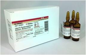
16w/v%多聚甲醛溶液,无甲醇(Methanol free)25%戊二醛溶液

16w/v%多聚甲醛溶液,无甲醇(Methanol free)
25%戊二醛溶液

  • 产品特性
  • 相关资料
  • Q&A
  • 参考文献

16w/v%多聚甲醛溶液,无甲醇(Methanol free)16w/v%多聚甲醛溶液,无甲醇(Methanol free)25%戊二醛溶液

25%戊二醛溶液

现出量少安瓿包装(10支装)

组织/细胞固定液

16w/v%多聚甲醛溶液,无甲醇(Methanol free)25%戊二醛溶液

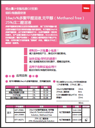
  多聚甲醛具有较高的反应性和细胞通透性,被广泛用作细胞中蛋白质和核酸的可逆交联剂和细胞/组织固定剂。关于配制16w/v%多聚甲醛溶液,由于多聚甲醛难溶于水,生成甲醛需要一定时间和操作。

  本产品是已配制16w/v%多聚甲醛溶液,为1mL和10mL的安瓿包装(惰性气体密封),无需担心储存期间由于氧化产生甲酸,任何时候打开使用都是“刚制成”状态。请配合25%戊二醛溶液一起使用。


◆优点

● 便利的一次性量小包装!

  减少废弃物的产生,降低对环境的影响。

● 加快实验/研究的速度!!
  减少溶液配制的时间和操作,提高实验效率。

● 适用于各种实验应用!!!

  能应对组织/细胞的各种实验。


◆应用实例

  用PBS等稀释16w/v%多聚甲醛溶液,无甲醇后使用。


应用

使用浓度

稀释

细胞/组织固定

4%

1:4

细胞/细胞器内交联

1%

1:16

ChIP化验

1%

1:16

流式细胞仪分析

2%

1:8


  可以使用Karnovsky,1965改良固定液(2%多聚甲醛、2.5%戊二醛)。


16w/v%多聚甲醛溶液

10mL

25%戊二醛溶液

8mL

0.2M 磷酸缓冲液(pH7.4)

40mL

蒸馏水

22mL


相关资料

16w/v%多聚甲醛溶液,无甲醇(Methanol free)25%戊二醛溶液

详情请点击图片


相关产品

产品编号

名称

规格

容量

071-02031

10%戊二醛溶液

10% Glutaraldehyde Solution

电子显微镜用

2ml×5A

072-02262

20% 戊二醛溶液

20% Glutaraldehyde Solution

电子显微镜用

25ml

076-02265

电子显微镜用

500ml

161-20141

4% 多聚甲醛溶液,磷酸盐缓冲溶液

4% Paraformaldehyde Phosphate Buffer Solution

组织固定用

100ml

163-20145

组织固定用

500ml

推荐原抗体产品

产品编号

名称

规格

容量

015-25191

抗磷酸化α-突触核蛋白,单克隆抗体(pSyn#64)

Anti Phosphorylated α-Synuclein, Monoclonal Antibody (pSyn#64)

免疫化学用

50μl

019-19741

抗Iba1,兔(免疫细胞化学用)

Anti Iba1, Rabbit (for Immunocytochemistry)

免疫化学用

50μg

014-24061

抗IDH1,单克隆抗体

Anti IDH1, Monoclonal Antibody

免疫化学用

100μg

011-24071

抗IDH2,单克隆抗体

Anti IDH2, Monoclonal Antibody

免疫化学用

100μg

018-24081

抗IDH1-R132H,单克隆抗体

Anti IDH1-R132H, Monoclonal Antibody

免疫化学用

100μg

015-24091

抗IDH1-R132S,单克隆抗体

Anti IDH1-R132S, Monoclonal Antibody

免疫化学用

100μg

018-24101

Anti-human Podoplanin,单克隆抗体

Anti HumanPodoplanin, Monoclonal Antibody

免疫化学用

100μg

015-24111

Anti mousepodoplanin,单克隆抗体

Anti mousePodoplanin, Monoclonal Antibody

免疫化学用

100μg

014-24821

抗CRMP2,单克隆抗体(9F)

Anti CRMP2, Monoclonal Antibody (9F)

免疫化学用

100μl

011-24831

抗CRMP5,单克隆抗体(KZ19)

Anti CRMP5, Monoclonal Antibody (KZ19)

免疫化学用

100μl

010-24801

抗磷酸化CRMP1/2(丝氨酸522),兔

Anti Phosphorylated CRMP1/2 (Ser522), Rabbit

免疫化学用

100μl


产品编号 产品名称 产品规格 产品等级 产品价格
077-06271 25% 戊二醛溶液 25% Glutaraldehyde Solution 10ml×10A 电子显微镜用
071-02031 10%戊二醛溶液 10% Glutaraldehyde Solution 2ml×5A 电子显微镜用
072-02262 20% 戊二醛溶液 20% Glutaraldehyde Solution 25ml 电子显微镜用
076-02265 20% 戊二醛溶液 20% Glutaraldehyde Solution 500ml 电子显微镜用
161-20141 4% 多聚甲醛溶液,磷酸盐缓冲溶液 4% Paraformaldehyde Phosphate Buffer Solution 100ml 组织固定用
163-20145 4% 多聚甲醛溶液,磷酸盐缓冲溶液 4% Paraformaldehyde Phosphate Buffer Solution 500ml 组织固定用
015-25191 抗磷酸化α-突触核蛋白,单克隆抗体(pSyn#64) Anti Phosphorylated α-Synuclein, 50μl 免疫化学用
019-19741 抗Iba1,兔(免疫细胞化学用) Anti Iba1, Rabbit (for Immunocytochemistry) 50μg 免疫化学用
014-24061 抗IDH1,单克隆抗体 Anti IDH1, Monoclonal Antibody 100μg 免疫化学用
011-24071 抗IDH2,单克隆抗体 Anti IDH2, Monoclonal Antibody 100μg 免疫化学用
018-24081 抗IDH1-R132H,单克隆抗体 Anti IDH1-R132H, Monoclonal Antibody 100μg 免疫化学用
015-24091 抗IDH1-R132S,单克隆抗体 Anti IDH1-R132S, Monoclonal Antibody 100μg 免疫化学用
018-24101 Anti-human Podoplanin,单克隆抗体 Anti HumanPodoplanin, Monoclonal Antibody 100μg 免疫化学用
015-24111 Anti mousepodoplanin,单克隆抗体 Anti mousePodoplanin, Monoclonal Antibody 100μg 免疫化学用
014-24821 抗CRMP2,单克隆抗体(9F) Anti CRMP2, Monoclonal Antibody (9F) 100μl 免疫化学用

免疫印迹显色试剂盒(抗小鼠)

免疫印迹显色试剂盒(抗小鼠)
Chromogenic Western Blot Kit (Anti-Mouse)

  • 产品特性
  • 相关资料
  • Q&A
  • 参考文献

免疫印迹显色试剂盒(抗小鼠)免疫印迹显色试剂盒(抗小鼠)

Chromogenic Western Blot Kit (Anti-Mouse)

信号、灵敏度更强的蛋白印迹显色试剂盒

Chromogenic Western Blot kit with increased signal and sensitivity

● 更灵敏的检测——能够检测较低浓度的蛋白质

● 易于使用——所有试剂均可随时使用或即可稀释,节省宝贵的时间

● 适应性好——适用于各种小鼠来源的蛋白质

显色蛋白质印迹试剂盒(抗小鼠)能通过免疫检测来识别蛋白质印迹上的一抗。该试剂盒内有完整的一套试剂,包含经优化的即用型或即可稀释试剂。

高灵敏度,可检测ng级蛋白,低表达蛋白无需纯化或浓缩样品,节省1~2h实验时间。


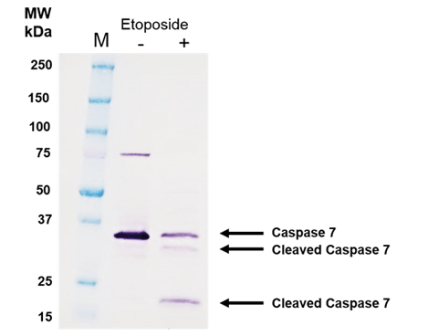
经过12.5 μM依托泊苷处理18 h的Jurkat细胞及未经处理对照的12 μg的总细胞裂解液上样


免疫印迹显色试剂盒(抗小鼠)

◆产品详情


反应时间:1小时

应用:免疫印迹法

物种反应性:小鼠

运输: 蓝冰

短期储存:+4°C

长期储存:+4°C

科学背景:免疫印迹法是一种常用的实验室技术,用于检测组织匀浆或提取物样品中的蛋白。由于具灵活性,它广

                 泛应用于分子生物学和生物化学,包括对翻译后修饰的检测以及对蛋白克隆的检验。根据蛋白质条带的

                 大小和颜色强度,即可对蛋白质进行半定量的估计。


◆试剂盒组成

1. Antibody Diluent, 4 x 90 mL
  用于稀释一抗和二抗的稀释液。


2. NBT/BCIP, 100 mL
  即用型印迹显色溶液。


3. Wash Buffer (10×), 2 x 100 mL

  用于洗涤膜的10×Wash Buffer。


4. Secondary Antibody (Anti-Mouse), 4.5 mL

  碱性磷酸酶偶联的抗小鼠二抗,用于在Western Blot中与小鼠源的一抗检测。


◆实验流程


试剂准备

二抗(抗小鼠)

在抗体阻断剂/稀释剂中以1:100-1000的稀释度制备二抗(抗小鼠)。例如,对于1:100的稀释度来说,将100 µL抗体加入到10 mL的抗体阻断剂/稀释剂中。


Wash Buffer(1×)

用90 mL去离子水稀释10 mL试剂盒中提供的10×Wash Buffer来制备1×Wash Buffer。 该缓冲液可以在室温下储存,可储存直至试剂盒有效期或储存3个月,以较早者为准。


◆实验流程

1. 在一抗孵育之后,将膜置于合适的塑料托盘中,在轨道式振荡器或摆动振荡器轻轻用1×Wash Buffer洗涤3-4

  次,每次10分钟(每次洗涤加入约10 mL的 1×Wash Buffer)。

2. 在最后一次洗涤期间,如前文所述制备二抗(抗-小鼠)。

3. 将带有二抗溶液的膜置于轨道式振荡器或摆动振荡器上适度摇荡,在室温下孵育1小时。

4. 在轨道式振荡器或摆动振荡器轻轻用1×Wash Buffer洗涤3-4次,每次10分钟

  (每次洗涤加入约10 mL的 1×Wash Buffer)。

5. 将膜置于黑暗中(用铝箔覆盖住托盘),加入NBT/BCIP(碱性磷酸酶显色试剂)孵育,直到膜上出现紫色条带

  (通常为10秒至2.5分钟)。期间不要摇动。

6. 在蒸馏水中洗涤3-5次。

7. 为获得最佳图像效果,请在第6步之后对膜进行扫描。

8. 如有必要,可使用滤纸或将膜置于红外灯下使膜干燥,并储存于4°C条件下。


◆产品列表

产品编号

产品名称

产品规格

ENZ-KIT182-0001

Chromogenic Western Blot Kit (Anti-Mouse)
免疫印迹显色试剂盒(抗小鼠)

1 Kit


◆相关产品

一抗来源并非小鼠?请用下面列表中的二抗替换本试剂盒中的二抗:

产品编号

产品名称

产品规格

ENZ-ACC103-0150

POLYVIEW® PLUS HRP (anti-rabbit) reagent
即用型HRP溶液(兔抗) 

150 test

ENZ-ACC104-0150

POLYVIEW® PLUS HRP (anti-mouse) reagent
即用型HRP溶液(鼠抗) 

150 test

ENZ-ACC110-0150

POLYVIEW® PLUS AP (anti-rabbit) reagent
POLYVIEW® +AP (抗兔)试剂 

150 test

ENZ-ACC114-0150

POLYVIEW® PLUS AP (anti-Mouse) Reagent
POLYVIEW® PLUS AP(抗老鼠)试剂 

150 test


想做多重检测?请看下面列表中的其他颜色的显色剂:

产品编号

产品名称

产品规格

ADI-950-140-0030

HIGHDEF red IHC chromogen (AP)
HighDef 红色免疫组化色原体(AP) 

30 mL

ADI-950-141-0030

HIGHDEF red IHC chromogen (AP, plus)
HighDef 红色免疫组化色原体(AP, plus) 

30 mL

ADI-950-150-0030

HIGHDEF blue IHC chromogen (AP)
HighDef 蓝色免疫组化色原体(AP) 

30 mL

ADI-950-151-0030

HIGHDEF blue IHC chromogen (HRP)
HighDef 蓝色免疫组化色原体(HRP) 

30 mL

ADI-950-170-0030

HIGHDEF yellow IHC chromogen (HRP)
HighDef 黄色免疫组化色原体 (HRP) 

30 mL

ADI-950-171-0030

HIGHDEF black IHC chromogen (HRP)
HighDef 黑色免疫组化色原体(HRP) 

30 mL

ADI-950-210-0030

HIGHDEF IHC chromogen substrate (AEC, stable)
HighDef 免疫组化色原体底物(AEC, 稳定) 

30 mL

ENZ-ACC130-0030

 HIGHDEF® Green AP Chromogen/Substrate
HIGHDEF® 绿AP染料/底物 

30 mL

产品编号 产品名称 产品规格 产品等级 产品价格

Fisher Scientific Traceable记忆计时器06-662-8-赛默飞中国代理商

产品信息
产品名称:
Fisher Scientific Traceable记忆计时器
产品型号:
06-662-8
Fisher Scientific Traceable记忆计时器06-662-8 产品特点
  Fisher Scientific Traceable记忆计时器技术参数/订购信息订货号FIS06-662-8特色带记忆功能Traceable证书有最长使用时间24小时通道数1通道分辨率1秒钟记忆功能有

Fisher Scientific Traceable记忆计时器06-662-8
产品详细信息:

Fisher Scientific Traceable记忆计时器

产品特色

使用 Traceable 记忆计时器,节约各项 测试的时间

● 这款独特的计时器能够倒数计时,配有闹 铃,并能保存设置记忆。用户可以按一分 钟的步进量,在 24 小时到 1 分钟的范围 内设置倒数计时。它的液晶显示数字高度 8.4 毫米,明亮清晰,以小时、分、秒的 格式显示时间(秒钟只在最后 10 分钟期间显示)。

● 用户只需碰触一只按钮,即可利用它独特的记忆功能,返回到先 前设定的时间显示。对它进行一次设置,即可保存记忆。许 多用户买好几只这款计时器,设定好记忆功能,然后将每一只计 时器专用于一项测试、水浴槽或仪表中。它响亮的闹铃声持续 60 秒钟,也可以提前手动关闭。它采用三键操作,使用简便。这 种袖珍型的装置配有夹子、磁体和底座,具备完全的灵活性。

● 记忆型计时器的石英晶体计时精度达 0.01%。为了确保精度,这 款计时器由 A2LA(A2LA 与 CNAL 的校准证书是互相承认的) 认可的 ISO 17025 校准实验室颁发了单独编号的 Traceable 证 书。这份证书表明,此项产品符合美国国家标准技术研究院(NIST) 颁布的标准。装置供货时配有可供 2 年使用的氧化银电池,电池 可以更换。装置尺寸是 60 毫米直径。

Fisher 长杆温度计15-078J-赛默飞中国代理商

产品信息
产品名称:
Fisher 长杆温度计
产品型号:
15-078J
Fisher 长杆温度计15-078J 产品特点
  Fisher 长杆温度计技术参数/订购信息订货号FIS15-078JFIS14-648-12FIS15-077-59FIS15-077-61Traceable证书有有有有量程-58至302℉/-50至150℃-58至572℉/-50至300℃精度&#177;1℃&#177;0.2℃&#177;1℃&#177;0.5℃探针长度203mm203mm289mm289mm装置尺寸21×279

Fisher 长杆温度计15-078J
产品详细信息:

Fisher Scientific Traceable长杆温度计

产品特色 取代玻璃汞柱温度计

● 这款温度计尺寸规格完美,适 合在烧瓶、比色杯、试管和烧 杯中使用,能够测量食品、气体、 样品、试剂、水浴槽和半固体 中的温度。它配备的不锈钢探 针能够耐受大多数实验室的化 学品,而且探针杆较长,使得 用户能够在比较深的容器中搅拌溶液的同时进行测量。它的量程 是 -58 到 572℉、-50 到 300℃,在 -20 到 200°之间的分辨 率为 0.1°(超出此范围则为 1°),在 -20 到 120℃之间的精 度为 ±1℃。UltraTM 型产品则在测试点达到 ±0.2 和 ±0.5℃的 精度。读数每秒钟刷新。 淘汰水银

● 这种产品采用塑料和不锈构的构造(不使用玻璃或水银),基本 上可以说是打不破的,这样就相当安全。所配高亮液晶显示屏高 度为 6.4 毫米。适合用于所有烘箱和孵化器。 精度符合国家标准技术研究院的规定

● 为了确保精度,这种温度计由 A2LA(A2LA 与 CNAL 的校准证 书是互相承认的)认可的 ISO 17025 校准实验室颁发了单独编号 的 Traceable 证书。这份证书表明,此项产品符合美国国家标准 技术研究院(NIST)颁布的标准。装置外壳采用丙烯腈-丁二烯苯乙烯共聚物(ABS)的耐用性塑料制成。其直径为 21 毫米, 杆直径3.5 毫米,重量不到29克,供货时配有可供使用1年的电池、 Traceable 证书及护套。

KBM581淋巴细胞无血清培养基1L装 美国Corning 货号:88-581-CM

名称:KBM581淋巴细胞无血清培养基1L装

品牌:美国Corning

订货号:88-581-CM

KBM581淋巴细胞无血清培养基1L装                                                        美国Corning                                                        货号:88-581-CM

咨询此产品

产品介绍

为了更好地服务中国不断发展的免疫治疗市场,康宁公司已经与日本Kohjin公司正式

签署协议,在免疫治疗产品领域进行合作。

    Kohjin总部位于日本东京,成立于1981年,拥有三十多年的细胞培养产品研发和生产

经验。Kohjin生产车间通过ISO13485认证,全部生产均遵循GMP标准,一直以来专注

于为市场提供技术领先、品质卓越的产品。

 KBM581淋巴细胞无血清培养基1L装                                                        美国Corning                                                        货号:88-581-CM

Corning KBM 581            

Ø KBM 551最新升级配方

Ø 专为CIK细胞培养开发

Ø 培养CIK细胞无需添加血清

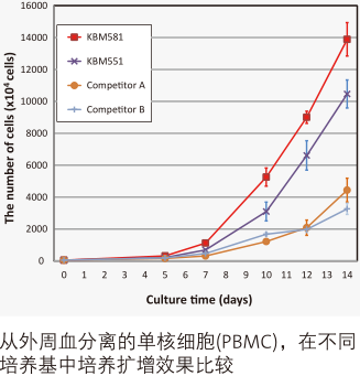
Ø 和市场上同类产品相比细胞增殖更快,活性细胞比例更高

KBM581

KBM551

Competitor A

Competitor B

CD3+/CD56+

13.00%

4.70%

8.80%

11.20%

CD3-/CD56+

2.30%

1.30%

1.10%

1.90%

CD3+/CD56-

84.60%

94.10%

90.00%

86.90%

CD3+/CD4+

25.80%

27.60%

24.40%

25.20%

CD3+/CD8+

66.10%

67.00%

62.10%

70.60%

   从外周血分离的单核细胞(PBMC),在不同培养基中培养后CIK细胞表型分析

  KBM581淋巴细胞无血清培养基1L装                                                        美国Corning                                                        货号:88-581-CM


产品用途

       此培养基适用于人外周血淋巴细胞的CIK诱导活化培养。 

     (需要添加IL-2、I N F – γ、抗C D 3 抗体及血清)


产品特点          

      ●用注射用水和多种药品级高纯度试剂配制而成的培养基。 

      ●蛋白质成份仅包括人血清白蛋白(药品级)和重组人胰岛素。 

      ●强化了培养基的缓冲能力,有效控制了pH值在最小范围内变动。 

      ●含有硫酸卡那霉素作为抗生素。 


注 意:本产品只适用于研究。 

           不含IL-2等细胞因子。 

形   状 :液体(方型PET瓶) 

容  量 :1000mL 

保存方法:避光保存(2-8℃) 

订购信息

货号

产品描述

体积

包装

88-581-CM

KBM581 淋巴细胞无血清培养基

1 升

Nuclease P1

Nuclease P1
核酸酶P1

  • 产品特性
  • 相关资料
  • Q&A
  • 参考文献

核酸酶P1Nuclease P1

Nuclease P1

基因研究用 for Genetic Research

制造商:FUJIFILM Wako Pure Chemical Corporation

储存条件:冷藏(冷藏运输)

CAS RN®54576-84-0


◆概况

核酸酶P1分解RNA和热变性DNA的3'-5'-磷酸二酯键。另外,核酸酶P1还分解单核寡核苷酸的3'-磷酸单酯键。如果的氯化钠(pH 6.0)含量超过400mmol/L,它不作用于双链DNA。

◆应用实例

A.

Sample : 200 μL of RNA or heat-denatured DNA at a density of 2 mg/mL.

B.

Reaction Buffer : 200 μL of 0.01 mol/L barbital(veronal) acetate buffer, pH 5.3*.

C.

Enzyme : 100 μL of 100 units/mL of Nucelase P1 solution with distilled water.

D.

top Solution : 500 μL of cold uranyl acetate buffer (0.25% of uranyl acetate and 2.5% perchloric acid)

*barbital (veronal) acetate buffer, pH 5.3: Prepare 0.01 mol/L Barbital Sodium Salt in water and adjust it to pH 5.3 with acetic acid.

1. Mix each 200 μL of A, B and C.

2. Incubate it at 50 degree C for 1 hour.

3. The reaction can be stopped by adding 500 μL of D.

◆参考文献

Masao Fujimoto, Akira Kuninaka & Hiroshi Yoshino(1973)Purification of a Nuclease from Penicillium citrinum, Agr. Biol. Chem.,38(4), 777-783.


◆用途

用于核酸相关的研究。

◆相关信息


外观:冻干品

来源:Penicillium citrinum

活性:约500 units/vial(3'-5'-磷酸二酯酶活性)

pH信息:最佳pH:5.3(RNA,热变性DNA),7.2(3'-AMP)

温度:最佳温度:约70°C,60°C(长时间孵育时)

 热稳定性:60°C以下(pH 5.3,30 min)稳定。

产品编号 产品名称 产品规格 产品等级 产品价格
145-08221 Nuclease P1
核酸酶P1
500 U 基因研究用

CYTO-ID® 自噬检测试剂盒

CYTO-ID® 自噬检测试剂盒
CYTO-ID® Autophagy detection kit

  • 产品特性
  • 相关资料
  • Q&A
  • 参考文献

CYTO-ID® 自噬检测试剂盒CYTO-ID® 自噬检测试剂盒

  CYTO-ID® Autophagy Detection Kit使用新型染料检测自噬小泡和监控活细胞自噬流,选择性标记积累的自噬小泡。染料已通过优化,不染溶酶体,在自噬前体、自噬体和自噬溶酶体里呈现出明亮的荧光。该试剂盒提供了一种无需细胞转染,可以在活细胞中监控细胞自噬的快速定量方法。

CYTO-ID® 自噬检测试剂盒

CYTO-ID® Autophagy Detection Kit

◆特点

  ● 无需转染,定量监测活细胞中的自噬情况

  ● 免去LC3-GFP转染所需的时间和精力以及转染效率的验证

  ● 含有特殊基团的专利染料选择性地染色自噬小泡

  ● 试剂盒中包含已知活性的自噬抑制剂与激活剂

  ● 快速量化原生异质性细胞群中的自噬

  ● 选择性地全方位染色,可检测和区分自噬流(autophagic flux)和自噬溶酶体的积累

  ● 不染溶酶体,减少其他染料的背景干扰

   便于高通量筛选自噬激活剂和抑制剂

◆原理


  该产品所用探针是阳离子两亲性示踪剂(CAT)染料,以类似诱导磷脂药物的方式迅速进入到细胞中。染料的功能基团能够选择性标记与自噬通路相关的小泡,而不会在溶酶体中聚集。


  胞内物质被扩大的膜囊包裹,吞噬泡形成双层膜囊泡,成为自噬体。自噬体外膜随后与溶酶体融合,和内部物质被自噬性溶酶体降解。自噬的各种调节因子也被描绘于图中。

CYTO-ID® 自噬检测试剂盒

自噬原理图

◆应用


省时省力,无需转染即可自快速全面的标记自噬小泡


  为了演示CYTO-ID® Green detection reagent的优势,先用RFP-LC3表达载体转染HeLa细胞, 10μM的Tamoxifen处理过夜,然后用CYTO-ID® 绿色检测试剂染色。LC3转染法需要过夜,而CYTO-ID® 绿色检测试剂的方法在15-30分钟内标记细胞达100%。图A:绿信号显示自噬小泡的CYTO-ID® 绿染;图B:成功转染细胞的RFP-LC3表达(红色);图C:自噬体的特异性标记LC3,与CYTO-ID® 绿色染料标记的小泡共定位的复合图像。

的发放

CYTO-ID® 自噬检测试剂盒


无转染的自噬检测


  Hela细胞进行饥饿与恢复,然后使用CYTO-ID® Green detection reagent标记。染料可以明确检测和量化自噬及与自噬诱导相关的自噬小泡前体。图1A:稳定表达GFP-LC3的CHO细胞,结果显示对照组与饥饿的细胞群的相对较差的基线分离,自噬难以量化。图1B:CYTO-ID® Autophagy Detection Kit特异性标记LC3蛋白依赖的自噬小泡,免去转染操作。

CYTO-ID® 自噬检测试剂盒


自噬积累和自噬流的可视化


  可用荧光显微镜观察CYTO-ID® 自噬绿色染料检测的自噬液泡积累和自噬流。Hela细胞用以下试剂进行模拟诱导,0.2% DMSO(A)和100μM  Clonidine hydrochloride(B)进行诱导,5μM Loperamide hydrochloride和1 μM PP242 hydrate(D)在37°C诱导12小时。处理后,细胞在37℃下用CYTO-ID® Green Detection reagent孵育10分钟,再用分析缓冲液清洗。细胞核被Hoechst 33342染料复染为蓝色。

CYTO-ID® 自噬检测试剂盒


避免非特异性溶酶体染色的背景干扰


  相比其他MDC染色溶酶体检测,CYTO-ID® 绿色染料无溶酶体染色背景,CYTO-ID® Autophagy kit无需紫外活细胞分析,在显微镜共标记应用中,与Hoechst染料兼容。

CYTO-ID® 自噬检测试剂盒


使用流式细胞仪通过CYTO-ID® Autophagy Detection Kit分析自噬


  对照组(红线峰值)无诱导,10 uM Tamoxifen (ALX-550-095)处理Jurkat细胞(白血病T细胞)(蓝线峰值)。处理18小时后,细胞结合CYTO-ID® 绿色检测试剂,然后不经过流式细胞仪清洗,进行分析。结果通过直方图呈现。对照组细胞被染色,但荧光亮度低。实验组中,CYTO-ID® 绿色荧光信号增加约2倍,表明Tamoxifen处理能引起Jurkat细胞中自噬的增加。


CYTO-ID® 自噬检测试剂盒

CYTO-ID® 自噬检测试剂盒

  使用mTOR激酶抑制剂Rapamycin处理HepG2细胞孵育过夜,结果显示CYTO-ID® 染色信号升高。

CYTO-ID® 自噬检测试剂盒

CYTO-ID® 绿色染料大多与RFP-LC3蛋白共定位。0.1μM Rapamycin(典型自噬诱导剂)处理过夜,转染Hela细胞表达RFP-LC3。A: CYTO-ID® Green染色; B: RFP-LC3; C: 合成图像。

CYTO-ID® 自噬检测试剂盒 2.0http://www.boppard.cn/product/show/735.html

◆相关产品

产品编号

产品名称

规格

备注

应用

ENZ-51002-25

GFP-Certified®Apoptosis/Necrosis detection kit

细胞凋亡/坏死检测试剂盒

25 assays

多重检测,区分正常、

早期凋亡、晚期凋亡和坏死细胞,与GFP和其他绿色荧光探针兼容。

FC,荧光显微镜,荧光检测

ENZ-51002-100

100 assays

ENZ-51021-K200

Nuclear-ID®Green

hromatin condensation detection kit

细胞核绿色染色体皱缩检测试剂盒


1 Ki

高渗透性的绿色荧光染色增强了细胞凋亡诱导染色质固缩。


FC,荧光显微镜,荧光检测

ENZ-52406

NUCLEAR-ID®Red DNA stain

DNA染色试剂盒(红色荧光)

200 µl

细胞可渗透的DNA染色应用广泛。

≥93%(HPLC),FC,荧光检测

ENZ-CHM103-0200

Nuclear-ID®Blue

DNA stain   (GFP-Certified®)

Nuclear   ID®蓝色DNA染色(GFP细胞系)

200 µ

细胞可渗透的DNA染色应用广泛。

≥93%(HPLC),FC,荧光检测

ENZ-51015-KP002

Lyso-ID®Red

cytotoxicity kit   (GFP-Certified®)

溶酶体细胞毒理检测试剂盒(红色荧光)(绿色细胞系)

1 Kit

快速,定量和HTS-兼容的检测活细胞毒性试剂。

荧光显微镜,荧光检测

ENZ-51035-0025

ProteoStat®Aggresome   etection kit

蛋白内稳态® 聚集体 检测 试剂盒

25 tests

Robust、定量的聚集小体用于神经退行性疾病,肝病和毒理学研究

FC,荧光显微镜,荧光检测

ENZ-51035-K100

100 tests


处理

目的

效果

μM

诱导时间 (hrs)

细胞系

饥饿

抑制哺乳动物Rapamycin (mTOR)

激活自噬

N/A

1~4

HeLa, HepG2, Jurkat

Rapamycin

抑制哺乳动物Rapamycin (mTOR)

激活自噬

0.2

6~18

HeLa, Jurkat

PP242

mTOR ATP-竞争性抑制剂

激活自噬

1

18

HeLa

Lithium

抑制IMPase 和降低inositol 和IP3水平; mTOR依赖性

激活自噬

10,000

18

HeLa, Jurkat

Trehalose

未知, mTOR依赖性

激活自噬

50,000

6

HeLa, Jurkat

Bafilomycin A1

抑制 Vacuolar-ATPase

抑制自噬

6~9*10-3

18

HeLa, Jurkat

Chloroquine

碱化 Lysosomal pH

抑制自噬

10~50

18

HeLa, Jurkat

Tamoxifen

增加细胞内 ceramide的水平和消除PI3K的抑制效果

激活自噬

4~10

6~18

HeLa, HepG2, Jurkat

Verapamil

钙离子通道阻滞剂;降低胞浆内Ca2+水平; mTOR依赖性

激活自噬

40~100

18

HeLa, Jurkat

HydroxyChloroquine

Alkalinizes Lysosomal pH

抑制自噬 

10

18

HeLa, Jurkat

Loperamide

Ca2+ 通道阻滞剂;降低胞浆内 Ca2+浓度; mTOR依赖性

激活自噬

5

18

HeLa

Clonidine

咪唑啉-1受体激动剂; 降低 cAMP 水平; mTOR依赖性

激活自噬

100

18

HeLa

MG-132

选择性蛋白酶抑制剂

激活自噬

2~5

18

HeLa, Jurkat

Norclomipramine

碱化Lysosomal pH

抑制自噬

5-20

18

HeLa

Epoxomicin

选择性蛋白酶抑制剂

诱导聚集体

0.5

18

HeLa

Velcade®

选择性蛋白酶抑制剂

诱导聚集体

0.5

18

HeLa

Amyloid beta peptide 1-42

引起氧化应激

诱导聚集体

25

18

SK-N-SH

Cyto-ID®检测试剂盒 Q&A


Q:Cyto-ID®自噬检测试剂与活细胞孵育的最长时间是多少(几分钟/几小时/几天)?
A:我们建议37℃孵育不超过30 分钟。很多实验表明,孵育1 小时不会伤害细胞。孵育超过1 小时,一些细胞看
上去状态不好。


Q:是否可用荧光显微镜观察3D 培养细胞的自噬?
A:我们没有用该染料染过3D 细胞,但是一小撮细胞是可以染色的。活体组织的话,由于染料的渗透性不好,不
能进入样本的内部,所以效果不理想。染白细胞是可以。


Q:用双重染料染在37℃孵育30 分钟后,细胞状态不好开始皱缩。有些细胞也从板上脱离,阳性对照和DMSO
对照没有观察到特异性染色。

A:对于比较敏感的细胞,优化流程如下:
1. 使用PBS/5%FBS 或者完全培养基作为assay buffer,最好不含酚红。
2. 如果细胞状态仍然不好,建议染色时间缩短至15~20 分钟。
3. 减少染料的浓度至1000×稀释。需要显微镜观察时延长荧光照射的时间。
4. 可能光褪色会导致信号模糊。建议使用封片剂防止光褪色。


Q:用流式检测的话,样品在用Cyto-ID®自噬检测试剂孵育后多久可用流式观察?操作手册是说30 分钟。是否可
以在检测之前短暂的将样品放置在冰上?

A:可以将样品放在冰上延长孵育的时间。一般的经验法则,尽可能缩短活细胞与染料的结合。缺氧,低温或者其
他应激状态也可以诱导自噬。


Q:GFP-CertifiedTM 细胞凋亡/坏死检测试剂盒(ENZ-51002),Cyto-ID®自噬检测试剂盒(ENZ-51031)是否可以
同时染色,检测细胞凋亡,坏死和自噬?

A:GFP-CertifiedTM 细胞凋亡/坏死检测试剂盒(ENZ-51002)和Cyto-ID®检测试剂盒(ENZ-51031)不能配合使
用,因为细胞凋亡和自噬检测的染料都发绿色荧光。这两个试剂盒可以平行使用。比如,用十字孢碱
(Staurosporine)处理细胞,一部分样品用GFP-CertifiedTM 细胞凋亡/坏死检测试剂盒检测,一部分样品用
Cyto-ID®检测试剂盒检测。


Q:Cyto-ID®自噬检测试剂盒(ENZ-51031) 的Cyto-ID®自噬检测试剂能否与ProteoStat®蛋白聚集检测
(ENZ-51023)的ProteoStat®检测试剂联合使用?

A:Cyto-ID®绿色自噬检测试剂与ProteoStat®检测试剂发的荧光颜色不同,他们可用于同一实验,客户需要优化
实验流程。

Q:Cyto-ID®自噬检测试剂的原理是什么?
A:染料是申请专利的。该染料是阳离子两性示踪剂(CAT)染料,跟很多阳离子化合物一样可以快速进入细胞。
染料穿过细胞膜双分子层后被动扩散,不需要蛋白结合或者转运子活性。可滴定部分的筛选使的染料不会再溶
酶体聚集,能够标记自噬通道相关的液泡。另外,染料的发射光密度增强后可以标记自噬液泡相关片层膜结构。
我们可以提供染料染自噬液泡的相关数据。
诱导自噬溶酶体,比如用氯喹或者baflimyocin 处理后,会导致荧光变亮(250~300%)。雷帕霉素诱导自
噬后能够用3-MA 抑制。在EBSS 中氨基酸缺乏会导致1 小时后在自噬液泡中染料的积累,这种积累是可逆的
(补充营养后即可恢复)。我们有一系列用于研究自噬的化合物(Screen-Well®Autophagy library)


Q:与单丹磺酰尸胺(MDC)相比,Cyto-ID®自噬检测试剂有哪些优势?
A:MDC 需要365nm UV 荧光照明,与通常流式配置的488n 激发源不兼容。Cyto-ID®自噬染料是488nm激发
波长,绿色的发射荧光,可以高亮自噬通道不同的液泡成分。需要注意的是,与lysomotrophic 染料不同,
MDC,LysoTrackerTMRed,吖啶橙主要是染溶酶体的,Cyto-ID®自噬染料只能染一点点溶酶体,主要是染自
噬溶酶体和早期自噬部分的选择标记物。


Q:与GFP-LC3 转染细胞相比,Cyto-ID®自噬检测试剂盒(ENZ-51031)的优势是什么?
A:Cyto-ID®检测试剂盒(ENZ-51031)的最大优势是不用转染细胞。转染很短暂,不同实验需要再购买相关试剂。
由于细胞过表达,会导致GFP-LC3 聚集,结果呈假阳性。转染效率低于100%,有些细胞不会表达必要的GFP
组件。该试剂盒检测的自噬,细胞群的一致性会更好。不需要每个细胞系都进行转染。也可以标记原代细胞。
原代细胞不容易进行转染。


Q:有哪些细胞系或者原代细胞可用Cyto-ID®自噬检测试剂?
A:用该试剂盒见检测过的代表性的细胞和细胞系包括肝细胞,SK-N-SH 神经瘤细胞,CHO 细胞,骨肉瘤来源细
胞,黑素留细胞,乳腺癌细胞,宫颈癌细胞,卵巢癌细胞,B 淋巴瘤细胞,结肠癌细胞,HepG 细胞,Jurkat T
细胞,牛大动脉内皮细胞(BAEC)。


Q:Cyto-ID®自噬检测试剂能否与其他染料配合使用,区分细胞核内体和自噬内涵体?
A:Cyto-ID®红色长期示踪剂染料(ENZ-51037)能够染细胞质膜,通过细胞内吞存在于细胞中。Cyto-ID®红色
染料与Cyto-ID®自噬检测试剂能够高亮异体吞噬vs 自噬。也可以Cyto-ID®自噬检测试剂与Lyso-ID®红色染
料或者LysoTrackerTM 红色染料进行活细胞染色,荧光显微镜观察。


Q:Cyto-ID®自噬检测试剂的稳定性如何?
A:荧光染料会发生光褪色,但是这种染料比荧光素更稳定。按照操作手册出来荧光染料。

Q:Cyto-ID®自噬检测试剂盒染色后是否可固定细胞?
A:在操作手册中提供了固定细胞的流程。不推荐用表面活性剂给细胞打孔,定位的荧光染料丢失。


Q:活细胞检测时,荧光信号强度可维持多长时间?
A:细胞诱导后发生自噬,在分析前染色。诱导时间少于1 小时或者诱导几天,诱导时间取决于自噬诱导剂。我们
没有在活细胞上进行长时间的染色,有客户发现可以至少染色24 小时。

参考文献

A comparison of strategies for immortalizing mouse embryonic fibroblasts: M.M. St. Amad, et al.; J. Biol. Methods 3, e41 (2016), Application(s): Detected autophagy induction in SV40 transformed versus serially passed MEFs, 全文

Astemizole-Histamine induces Beclin-1-independent autophagy by targeting p53-dependent crosstalk between autophagy and apoptosis: R. Jakhar, et al.; Cancer Lett. 372, 89 (2016), Application(s): Flow cytometry analysis, 摘要;

Atg5 Is Essential for the Development and Survival of Innate Lymphocytes: T.E. O'Sullivan, et al.; Cell Rep. 15, 1910 (2016), Application(s): Liver lymphocytes were harvested and stained with cell surface antibodies and then incubated with 1:400 Cyto-ID Autophagy Detection Reagent, 摘要全文

Autophagy-related gene 5 and Wnt5 signaling pathway requires differentiation of embryonic stem cells into odontoblast-like cells: N. Ozeki, et al.; Exp. Cell Res. 341, 92 (2016), Application(s): Autophagy flux, 摘要;

DHA-induced stress response in human colon cancer cells-focus on oxidative stress and autophagy: K. Pettersen, et al.; Free Radic. Biol. Med. 90, 158 (2016), Application(s):Autophagy determined by flow cytometry, 摘要;

Diosgenin induces ROS-dependent autophagy and cytotoxicity via mTOR signaling pathway in chronic myeloid leukemia cells: S. Jiang, et al.; Phytomedicine 23, 243 (2016),Application(s): Confocal immunofluorescence, 摘要;

Efavirenz causes oxidative stress, endoplasmic reticulum stress, and autophagy in endothelial cells: M. Weiss, et al.; Cardiovasc. Toxicol. 16, 90 (2016), 摘要;

MiR-193b promotes autophagy and non-apoptotic cell death in oesophageal cancer cells: M.J. Nyhan, et al.; BMC Cancer 16, 101 (2016), Application(s): Assay used to stain live cells, 摘要全文

(Pro)renin receptor regulates autophagy and apoptosis in podocytes exposed to high glucose: C. Li, et al.; Am. J. Physiol. Endocrinol. Metab. 309, E302 (2015), Application(s):Confocal microscopy using mouse podocytes, 摘要;

A rapid and high content assay that measures cyto-ID-stained autophagic compartments and estimates autophagy flux with potential clinical applications: S. Guo, et al.; Autophagy11, 560 (2015), Application(s): Fluorescent detection, Microplate Reader , 摘要全文

A Systems Approach Identifies Essential FOXO3 Functions at Key Steps of Terminal Erythropoiesis: R. Liang, et al.; PLoS Genet. 11, e1005526 (2015), Application(s):Autophagy flux measured by flow cytometry , 摘要全文

ABT-888 enhances cytotoxic effects of temozolomide independent of MGMT status in serum free cultured glioma cells: R.K. Balvers, et al.; J. Transl. Med. 13, 74 (2015),Application(s): Assay, 摘要全文

Activation of autophagy in response to nanosecond pulsed electric field exposure: J.C. Ullery, et al.; Biochem. Biophys. Res. Commun. 458, 411 (2015), Application(s):Fluorescence microscopy using U937 monocyte and CHO-K1 cell lines, 摘要;

Aflatoxin biosynthesis is a novel source of reactive oxygen species-a potential redox signal to initiate resistance to oxidative stress?: L.V. Roze, et al.; Toxins (Basel). 7, 1411 (2015),Application(s): Assay, 摘要全文

Alisertib induces cell cycle arrest and autophagy and suppresses epithelial-to-mesenchymal transition involving PI3K/Akt/mTOR and sirtuin 1-mediated signaling pathways in human pancreatic cancer cells: F. Wang, et al.; Drug Des. Devel. Ther. 9, 575 (2015), Application(s): Flow cytometry using PANC-1 and BxPC-3 pancreatic cancer cell lines, 摘要全文

Alisertib, an Aurora kinase A inhibitor, induces apoptosis and autophagy but inhibits epithelial to mesenchymal transition in human epithelial ovarian cancer: Y.H. Ding, et al.; Drug Des. Devel. Ther. 9, 425 (2015), Application(s): Flow cytometry using SKOV3 and OVCAR-4 ovarian cancer cell lines, 摘要全文

Andrographolide Analogue Induces Apoptosis and Autophagy Mediated Cell Death in U937 Cells by Inhibition of PI3K/Akt/mTOR Pathway: D. Kumar, et al.; PLoS One 10, e0139657 (2015), Application(s): Flow cytometric analysis of Cyto-ID Green Detection Reagent , 摘要全文

Apoptotic Cell Death Induced by Resveratrol Is Partially Mediated by the Autophagy Pathway in Human Ovarian Cancer Cells: F. Lang, et al.; PLoS One 10, e0129196 (2015),Application(s): Live Cell Imaging, 摘要全文

Araguspongine C induces autophagic death in breast cancer cells through suppression of c-Met and HER2 receptor tyrosine kinase signaling: M.R. Akl, et al.; Mar. Drugs 13, 288 (2015), Application(s): Flow cytometry using BT-474 breast cancer cell line, 摘要全文

Autocrine VEGF maintains endothelial survival through regulation of metabolism and autophagy: C.K. Domigan, et al.; J. Cell. Sci. 128, 2236 (2015), 摘要;

Autophagy is activated in systemic lupus erythematosus and required for plasmablast development: A.J. Clarke, et al.; Ann. Rheum. Dis. 74, 912 (2015), 摘要全文

Autophagy limits proliferation and glycolytic metabolism in acute myeloid leukemia: A.S. Watson, et al.; Cell Death Discov. 1, 15008 (2015), Application(s): CytoID assay in human and mouse HSCs, 摘要全文

Baicalin inhibits autophagy induced by influenza A virus H3N2: H.Y. Zhu, et al.; Antiviral Res. 113, 62 (2015), Application(s): Fluorescence microscopy using A549 human lung cancer cell line, 摘要;

Bardoxolone methyl induces apoptosis and autophagy and inhibits epithelial-to-mesenchymal transition and stemness in esophageal squamous cancer cells: Y.Y. Wang, et al.; Drug Des. Devel. Ther. 9, 993 (2015), Application(s): Flow Cytometry, 摘要全文

Cell-penetrating peptide derived from human eosinophil cationic protein inhibits mite allergen Der p 2 induced inflammasome activation: S.J. Yu, et al.; PLoS One 10, e0121393 (2015), Application(s): Flow cytometry of THP-1 leukemia cell line, 摘要全文

Chemoproteomics Reveals Novel Protein and Lipid Kinase Targets of Clinical CDK4/6 Inhibitors in Lung Cancer: N.J. Sumi, et al.; ACS Chem. Biol. 10, 2680 (2015),Application(s): Quantification of autophagosomes, 摘要;

Circulating hemocytes from larvae of the Japanese rhinoceros beetle Allomyrina dichotoma (Linnaeus) (Coleoptera: Scarabaeidae) and the cellular immune response to microorganisms: S. Hwang, et al.; PLoS One 10, e0128519 (2015), Application(s):Fluorescence microscopy using hemocytes from Japanese rhinoceros beetle Allomyrina dichotoma larvae, 摘要全文

Citreoviridin induces ROS-dependent autophagic cell death in human liver HepG2 cells: Y.N. Liu, et al.; Toxicon. 95, 30 (2015), Application(s): Fluorescence microscopy using HepG2 cell line, 摘要;

Clozapine induces autophagic cell death in non-small cell lung cancer cells: Y.C. Yin, et al.; Cell. Physiol. Biochem. 35, 945 (2015), 摘要;

Coffee and caffeine potentiate the antiamyloidogenic activity of melatonin via inhibition of Aβ oligomerization and modulation of the Tau-mediated pathway in N2a/APP cells: L.F. Zhang, et al.; Drug Des. Devel. Ther. 9, 241 (2015), Application(s): Flow Cytometry,摘要全文

Combination of the mTOR inhibitor RAD001 with temozolomide and radiation effectively inhibits the growth of glioblastoma cells in culture: H. Burckel, et al.; Oncol. Rep. 33, 471 (2015), 摘要;

Danusertib Induces Apoptosis, Cell Cycle Arrest, and Autophagy but Inhibits Epithelial to Mesenchymal Transition Involving PI3K/Akt/mTOR Signaling Pathway in Human Ovarian Cancer Cells: D. Zi, et al.; Int. J. Mol. Sci. 16, 27228 (2015), Application(s): Confocal fluorescence microscopy, 摘要全文

Danusertib, a potent pan-Aurora kinase and ABL kinase inhibitor, induces cell cycle arrest and programmed cell death and inhibits epithelial to mesenchymal transition involving the PI3K/Akt/mTOR-mediated signaling pathway in human gastric cancer AGS and NCI-N78 cells: C.X. Yuan, et al.; Drug Des. Devel. Ther. 9, 1293 (2015), Application(s): Flow cytometry using AGS and NCI-N78 gastric cancer cell lines, 摘要全文

Defective autophagy in vascular smooth muscle cells alters contractility and Ca²⁺ homeostasis in mice: C.F. Michiels, et al.; Am. J. Physiol. Heart Circ. Physiol. 308, H557 (2015), 摘要;

Effects of cyclodextrins on GM1‐gangliosides in fibroblasts from GM1‐gangliosidosis patients: Y. Maeda, et al.; J. Pharm. Pharmacol. 67, 1133 (2015), 摘要;

Endurance exercise training induces fat depot-specific differences in basal autophagic activity: G. Tanaka, et al.; Biochem. Biophys. Res. Commun. 466, 512 (2015),Application(s): Detect the formation of autophagosomes, 摘要;

Erbin is a novel substrate of the Sag-βTrCP E3 ligase that regulates KrasG12D-induced skin tumorigenesis: C.M. Xie, et al.; J. Cell. Biol. 209, 721 (2015), 摘要;

Evaluation of Antitumor Effects of Folate-Conjugated Methyl-β-cyclodextrin in Melanoma: K. Motoyama, et al.; Biol. Pharm. Bull. 38, 374 (2015), Application(s): Fluorescence Microscopy, 摘要全文

Exchange protein directly activated by cAMP 1 promotes autophagy during cardiomyocyte hypertrophy: A.C. Laurent, et al.; Cardiovasc. Res. 105, 55 (2015), Application(s):Fluorescence microscopy using rat neonatal ventricular myocytes, 摘要;

Glutathione-S-transferase omega 1 (GSTO1-1) acts as mediator of signaling pathways involved in aflatoxin B1-induced apoptosis-autophagy crosstalk in macrophages: S. Paul, et al.; Free Radic. Biol. Med. 89, 1218 (2015), Application(s): Determination of autophagy with immunocytochemistry , 摘要;

GMI, an immunomodulatory protein from Ganoderma microsporum, potentiates cisplatin-induced apoptosis via autophagy in lung cancer cells: I.L. Hsin, et al.; Mol. Pharm. 12, 1534 (2015), 摘要;

Induction of apoptosis and autophagy via sirtuin1- and PI3K/Akt/mTOR-mediated pathways by plumbagin in human prostate cancer cells: Z.W. Zhou, et al.; Drug Des. Devel. Ther. 9, 1511 (2015), Application(s): Assay, 摘要全文

Induction of autophagy is a key component of all-trans-retinoic acid-induced differentiation in leukemia cells and a potential target for pharmacologic modulation: N. Orfali, et al.; Exp. Hematol. 43, 781 (2015), Application(s): Flow cytometry analysis of NB4 and HL60 promyelocytic leukemia cell lines, 摘要;

Inhibition of Autophagy Potentiated the Antitumor Effect of Nedaplatin in Cisplatin-Resistant Nasopharyngeal Carcinoma Cells: Z. Liu, et al. ; PLoS One 10, e0135236 (2015),Application(s): Cell culture, 摘要全文

Inhibition of mitotic Aurora kinase A by alisertib induces apoptosis and autophagy of human gastric cancer AGS and NCI-N78 cells: C.X. Yuan, et al.; Drug Des. Devel. Ther. 9, 487 (2015), Application(s): Flow cytometry using AGS and NCI-N78 gastric cancer cell lines, 摘要全文

Interferon Regulatory Factor-1 signaling regulates the switch between autophagy and apoptosis to determine breast cancer cell fate: J.L. Schwartz-Roberts, et al.; Cancer Res.75, 1046 (2015), 摘要;

Interplay of Oxidative Stress and Autophagy in PAMAM Dendrimers-Induced Neuronal Cell Death : Y. Li, et al.; Theranostics 5, 1363 (2015), Application(s): Confocal fluorescence assay, 摘要全文

Invariant NKT cells require autophagy to coordinate proliferation and survival signals during differentiation: B. Pei, et al.; J. Immunol. 194, 5872 (2015), 摘要;

Involvement of fish signal transducer and activator of transcription 3 (STAT3) in nodavirus infection induced cell death: Y. Huang, et al.; Fish Shellfish Immunol. 43, 241 (2015),Application(s): Fluorescence microscopy of Grouper (fish) brain cells, 摘要;

Is the autophagy a friend or foe in the silver nanoparticles associated radiotherapy for glioma?: H. Wu, et al.; Biomaterials 62, 47 (2015), Application(s): Fluorescence microscopy using U251 human glioma cell line, 摘要;

Kaposi's sarcoma-associated herpesvirus induces Nrf2 activation in latently infected endothelial cells through SQSTM1 phosphorylation and interaction with polyubiquitinated Keap1: O. Gjyshi, et al.; J. Virol. 89, 2268 (2015), 摘要;

KLF4-SQSTM1/p62-associated prosurvival autophagy contributes to carfilzomib resistance in multiple myeloma models: I. Riz, et al.; Oncotarget 6, 17814 (2015), Application(s):FACS, 摘要全文

Lithium modulates autophagy in esophageal and colorectal cancer cells and enhances the efficacy of therapeutic agents in vitro and in vivo: T.R. O'Donovan, et al.; PLoS One 10, e0134676 (2015), Application(s): Flow cytometry analysis using human esophageal and murine colon cancer cell lines, 摘要全文

Methicillin-Resistant Staphylococcus aureus Adaptation to Human Keratinocytes: G. Soong, et al.; MBio. 6, e00289-15 (2015), Application(s): Assay, 摘要全文

Mevalonate pathway regulates cell size homeostasis and proteostasis through autophagy: T.P. Miettinen, et al.; Cell Rep. 13, 2610 (2015), Application(s): Flow cytometry analysis of autophagy using Jurkat, U2OS, Kc167 and HUVEC cells, 摘要;

MiR-29b replacement inhibits proteasomes and disrupts aggresome+autophagosome formation to enhance the antimyeloma benefit of bortezomib: S. Jagannathan, et al.; Leukemia 29, 727 (2015), Application(s): Detection of autophagy by fluorescence microscopy in multiple myeloma cell lines, 摘要全文

Molecular chaperone GRP78 enhances aggresome delivery to autophagosomes to promote drug resistance in multiple myeloma: M.A. Abdel Malek, et al.; Oncotarget 6, 3098 (2015), Application(s): Confocal Microscopy, 摘要全文

Molecular cloning and characterization of autophagy-related gene TmATG8 in Listeria-invaded hemocytes of Tenebrio molitor: H. Tindwa, et al.; Dev. Comp. Immunol. 51, 88 (2015), Application(s): Fluorescence microscopy using hemocytes from Tenebrio molitor larvae, 摘要;

Molecular pathway of near-infrared laser phototoxicity involves ATF-4 orchestrated ER stress: I. Khan, et al.; Sci. Rep. 5, 10581 (2015), Application(s): Fluorescence microscopy autophagy assay, 摘要全文

N-Myc and STAT Interactor regulates autophagy and chemosensitivity in breast cancer cells: B.J. Metge, et al.; Sci. Rep. 5, 11995 (2015), Application(s): Fluorescent detection,摘要全文

Novel autophagy inducers lentztrehaloses A, B and C: S.I. Wada, et al.; J. Antibiot. (Tokyo)68, 521 (2015), Application(s): Fluorescence microscopy using Mewo melanoma and OVK18 ovarian cancer cell lines, 摘要;

Novel small-molecule SIRT1 inhibitors induce cell death in adult T-cell leukaemia cells: T. Kozako, et al.; Sci. Rep. 5, 11345 (2015), Application(s): Flow cytometry using a variety of cancer cell lines, 摘要全文

Novel targeting of PEGylated liposomes for codelivery of TGF-β1 siRNA and four antitubercular drugs to human macrophages for the treatment of mycobacterial infection: a quantitative proteomic study: N. Niu, et al. ; Drug Des. Devel. Ther. 9, 4441 (2015),Application(s): Autophagy of human macrophages by flow cytometry, 摘要;

Paraptosis cell death induction by the thiamine analog benfotiamine in leukemia cells: N. Sugimori, et al.; PLoS One 10, e0120709 (2015), Application(s): Flow cytometry using HL60 leukemia cell line, 摘要全文

Plumbagin induces G2/M arrest, apoptosis, and autophagy via p38 MAPK- and PI3K/Akt/mTOR-mediated pathways in human tongue squamous cell carcinoma cells: S.T. Pan, et al.; Drug Des. Devel. Ther. 9, 1601 (2015), Application(s): Assay, 摘要全文

Pro-apoptotic and pro-autophagic effects of the Aurora kinase A inhibitor alisertib (MLN8237) on human osteosarcoma U-2 OS and MG-63 cells through the activation of mitochondria-mediated pathway and inhibition of p38 MAPK/PI3K/Akt/mTOR signaling pathway: N.K. Niu, et al.; Drug Des. Devel. Ther. 9, 1555 (2015), Application(s): Assay,摘要全文

Reduced FoxO3a expression causes low autophagy in idiopathic pulmonary fibrosis fibroblasts on collagen matrix: J. Im, et al.; Am. J. Physiol. Lung Cell. Mol. Physiol. 309, L552 (2015), 摘要;

S-Adenosyl-L-methionine-competitive inhibitors of the histone methyltransferase EZH2 induce autophagy and enhance drug sensitivity in cancer cells: T.P. Liu, et al.; Anticancer Drugs 26, 139 (2015), Application(s): Fluorescence microscopy using MDA-MB-231 breast cancer cell line, 摘要全文

Schisandrin B inhibits cell growth and induces cellular apoptosis and autophagy in mouse hepatocytes and macrophages: implications for its hepatotoxicity: Y. Zhang, et al.; Drug Des. Devel. Ther. 9, 2001 (2015), Application(s): Flow cytometry using AML-12 hepatocyte and RAW 264.7 leukemia cell lines, 摘要全文

Src/STAT3-dependent heme oxygenase-1 induction mediates chemoresistance of breast cancer cells to doxorubicin by promoting autophagy: Q. Tan, et al.; Cancer Sci. 106, 1023 (2015), 摘要;

The CCL2 chemokine is a negative regulator of autophagy and necrosis in luminal B breast cancer cells: W.B. Fang, et al.; Breast Cancer Res. Treat. 150, 309 (2015), 摘要;

The investigational Aurora kinase A inhibitor alisertib (MLN8237) induces cell cycle G2/M arrest, apoptosis, and autophagy via p38 MAPK and Akt/mTOR signaling pathways in human breast cancer cells: J.P. Li, et al.; Drug Des. Devel. Ther. 9, 1627 (2015),Application(s): Assay, 摘要全文

The pan-inhibitor of Aurora kinases danusertib induces apoptosis and autophagy and suppresses epithelial-to-mesenchymal transition in human breast cancer cells: J.P. Li, et al.; Drug Des. Devel. Ther. 9, 1027 (2015), Application(s): Assay, 摘要全文

The role of autophagy in the cytotoxicity induced by recombinant human arginase in laryngeal squamous cell carcinoma: C. Lin, et al.; Appl. Microbiol. Biotechnol. 99, 8487 (2015), 摘要;

A Novel CXCR3-B Chemokine Receptor-induced Growth-inhibitory Signal in Cancer Cells Is Mediated through the Regulation of Bach-1 Protein and Nrf2 Protein Nuclear Translocation : M. Balan & S. Pal; J. Biol. Chem. 289, 3126 (2014), Application(s): Monitor autophagy in MCF-7 and T47D breast cancer cells by flow cytometry and fluorescence microscopy, 摘要;

Adaptive responses to glucose restriction enhance cell survival, antioxidant capability, and autophagy of the protozoan parasite Trichomonas vaginalis: K.Y. Huang, et al.; Biochim. Biophys. Acta. 1840, 53 (2014), 摘要;

Autophagy in the brain of neonates following hypoxia-ischemia shows sex-and region-specific effects: S.N. Weis, et al.; Neuroscience 256, 201 (2014), 摘要;

Cannabinoid-induced autophagy regulates suppressor of cytokine signaling-3 in intestinal epithelium: L.C. Koay, et al.; Am. J. Physiol. Gastrointest. Liver Physiol. 307, G140 (2014),Application(s): Detection of autophagy in human colonic epithelial cell line Caco-2 by Confocal imaging, 摘要全文

Caveolin-1 Is a Critical Determinant of Autophagy, Metabolic Switching, and Oxidative Stress in Vascular Endothelium: T. Shiroto, et al.; PLoS One 9, e87871 (2014), 摘要;全文

Connective tissue diseases: How do autoreactive B cells survive in SLE-autophagy?: N.J. Bernard; Nat. Rev. Rheumatol. 10, 128 (2014), (Review), 摘要;

Defective Autophagosome Trafficking Contributes to Impaired Autophagic Flux in Coronary Arterial Myocytes Lacking CD38 Gene: Y. Zhang, et al.; Cardiovasc. Res. 102, 68 (2014),摘要;

Defects in mitochondrial clearance predispose human monocytes to interleukin-1β hyper-secretion: R. van der Burgh, et al.; J. Biol. Chem. 289, 5000 (2014), 摘要全文

Early biomarkers of response to carfilzomib in multiple myeloma (MM): Modulation of CXCR4 and induction of autophagy: M. Bhutani, et al.; J. Clin. Oncol. 32, e19572 (2014),Application(s): Quantification of autophagy in malignant plasma cells from bone marrow aspirates by flow cytometry with the Cyto-ID autophagy detection kit,

Enhancement of dynein-mediated autophagosome trafficking and autophagy maturation by ROS in mouse coronary arterial myocytes: M. Xu, et al.; J. Cell. Mol. Med. 18, 2165 (2014), 摘要全文

Flow Cytometric Analysis of Autophagic Activity with Cyto-ID Staining in Primary Cells: M. Stankov, et al.; Bio-Protocol (2014), Application(s): FC in primary BMDCs, 全文

High-Content Assays for Hepatotoxicity Using Induced Pluripotent Stem Cell-Derived Cells: O. Sirenko, et al.; Assay Drug Dev. Technol. 12, 43 (2014), 摘要全文

Histone deacetylase inhibitors induce apoptosis in myeloid leukemia by suppressing autophagy: M.V. Stankov, et al.; Leukemia 28, 577 (2014), 摘要;

Histone deacetylase inhibitors potentiate VSV oncolysis in prostate cancer cells by modulating NF-κB dependent autophagy: L. Shulak, et al.; J. Virol. 88, 2927 (2014),摘要;

In vitro and in vivo characterization of porcine acellular dermal matrix for gingival augmentation procedures: A.M. Pabst, et al.; J. Periodontal. Res. 49, 371 (2014), 摘要;

Inhibition of Autophagic Flux by Salinomycin Results in Anti-Cancer Effect in Hepatocellular Carcinoma Cells: J. Klose, et al.; PLoS One 9, e95970 (2014),Application(s): Autophagy detection in human hepatocellular carcinoma , 摘要全文

Inhibition of stress induced premature senescence in presenilin-1 mutated cells with water soluble Coenzyme Q10: D. Ma, et al.; Mitochondrion 17C, 106 (2014), Application(s):Autophagic vacuoles in Alzheimer's Disease fibroblasts detected with CytoID® Green Autophagy Detection kit, 摘要;

Involvement of autophagy in recombinant human arginase-induced cell apoptosis and growth inhibition of malignant melanoma cells: Z. Wang, et al.; Appl. Microbiol. Biotechnol.98, 2485 (2014), 摘要;

MiR-216a: a link between endothelial dysfunction and autophagy: R. Menghini, et al.; Cell Death Dis. 5, e1029 (2014), 摘要;

Novel estradiol analogue induces apoptosis and autophagy in esophageal carcinoma cells: E. Wolmarans, et al.; Cell. Mol. Biol. Lett. 19 , 98 (2014), Application(s): Autophagy detection in esophageal carcinoma SNO cell , 摘要;

Novel sorafenib-based structural analogues: in-vitro anticancer evaluation of t-MTUCB and t-AUCMB: A.T. Wecksler, et al.; Anticancer Drugs 25, 433 (2014), 摘要;

Photodynamic therapy with the novel photosensitizer chlorophyllin f induces apoptosis and autophagy in human bladder cancer cells: D. Lihuan, et al.; Lasers Surg. Med. 46, 319 (2014), 摘要;

Plumbagin induces apoptotic and autophagic cell death through inhibition of the PI3K/Akt/mTOR pathway in human non-small cell lung cancer cells: Y.C.Li, et al.; Cancer Lett. 344, 239 (2014), 摘要;

Potential of adenovirus-mediated REIC/Dkk-3 gene therapy for use in the treatment of pancreatic cancer: D. Uchida, et al.; J. Gastroenterol. Hepatol. 29, 973 (2014), 摘要;

Sirt1 modulates endoplasmic reticulum stress-induced autophagy in heart: A. Guilbert, et al.; Cardiovasc. Res. 103 (suppl 1), S13 (2014), Application(s): Evaluation of Autophagy in H9c2 cells, rat cardiomyoblasts by flow cytometry, 全文

STAT3 down regulates LC3 to inhibit autophagy and pancreatic cancer cell growth: J. Gong, et al.; Oncotarget 5, 2529 (2014), Application(s): Autophagic vacuole formation was detected by microscopy and autophagosome formation was determined by flow cytometry in human pancreatic cancer cells Capan-2, 摘要全文

T-Cell Autophagy Deficiency Increases Mortality and Suppresses Immune Responses after Sepsis: C.W. Lin, et al.; PLoS One 9, e102066 (2014), Application(s): Quantification of autophagosomes and autolysosomes staining in CD4+ and CD8+ cell population by flow cytometry , 摘要全文

Tetracyclines cause cell stress-dependent ATF4 activation and mTOR inhibition: A. Brüning, et al.; Exp. Cell Res. 320, 281 (2014), 摘要;

The core autophagy protein ATG4B is a potential biomarker and therapeutic target in CML stem/progenitor cells: K. Rothe, et al.; Blood 123, 3622 (2014), Application(s): Monitor autophagy flux in hematopoietic stem/progenitor cells, 摘要;

Androgen deprivation and androgen receptor competition by bicalutamide induce autophagy of hormone-resistant prostate cancer cells and confer resistance to apoptosis: B. Boutin, et al.; Prostate 73, 1090 (2013), Application(s): Measurement of autophagic flux in prostate cancer cells, 摘要;

Arenobufagin, a natural bufadienolide from toad vonem, induces apoptosis and autophagy in human hepatocellular carcinoma cells through inhibition of PI3K/Akt/mTOR pathway: D.M. Zhang, et al.; Carcinogenesis 34, 1331 (2013), Application(s): Autophagy detection in hepatocellular carcinoma, 摘要;

Autophagy Plays a Critical Role in ChLym-1-Induced Cytotoxicity of Non-Hodgkin's Lymphoma Cells: J. Fan, et al.; PLoS One. 8, e72478 (2013), 摘要全文

BCL-2 inhibitors sensitize therapy-resistant chronic lymphocytic leukemia cells to VSV oncolysis: S. Samuel, et al.; Mol. Ther. 21, 1413 (2013), 摘要;

Bleomycin exerts ambivalent antitumor immune effect by triggering both immunogenic cell death and proliferation of regulatory T cells: H. Bugaut, et al.; PLoS One 8, e65181 (2013),Application(s): Measurement of autophagy by flow cytometry and fluorescence microscopy, 摘要全文

Celecoxib enhances radiosensitivity of hypoxic glioblastoma cells through endoplasmic reticulum stress: K. Suzuki, et al.; Neuro. Oncol. 15, 1186 (2013), 摘要;

Chloroquine Engages the Immune System to Eradicate Irradiated Breast Tumors in Mice: J.A. Ratikan, et al.; Int. J. Radiat. Oncol. Biol. Phys. 87, 761 (2013), 摘要;

Dietary Resveratrol Prevents Development of High-Grade Prostatic Intraepithelial Neoplastic Lesions: Involvement of SIRT1/S6K Axis: G. Li, et al.; Cancer Prev. Res 6, 27 (2013), Application(s): Effects of Resveratrol on prostate tumorigenesis, 摘要;

Enhancement of autophagy by simvastatin through inhibition of Rac1-mTOR signaling pathway in coronary arterial myocytes: Y.M. Wei, et al.; Cell. Physiol. Biochem. 31, 925 (2013), 摘要全文

GX15-070 (obatoclax) induces apoptosis and inhibits cathepsin D and L mediated autophagosomal lysis in antiestrogen resistant breast cancer cells: J.L. Schwartz-Roberts, et al.; Mol. Cancer Ther. 12, 448 (2013), Application(s): Autophagy detection in breast cancer cells, 摘要;

Hydroxychloroquine preferentially induces apoptosis of CD45RO+ effector T cells by inhibiting autophagy: A possible mechanism for therapeutic modulation of T cells: J. van Loodregt, et al.; J. Allergy Clin. Immunol. 131, 1443 (2013), Application(s): Detection of autophagy in CD4+ T cells and PBMC by flow cytometry , 摘要全文

Interactions between autophagic and endo-lysosomal markers in endothelial cells: C.L. Oeste, et al.; Histochem. Cell. Biol. 139, 659 (2013), 摘要;

Involvement of cholesterol depletion from lipid rafts in apoptosis induced by methyl-β-cyclodextrin: R. Onodera, et al.; Int. J. Pharm. 452, 116 (2013), Application(s):Measurement of autophagy by fluorescence microscopy, 摘要;

ISG15 deregulates autophagy in genotoxin-treated ataxia telangiectasia cells: S.D. Desai, et al.; J. Biol. Chem. 288, 2388 (2013), Application(s): Fluorescence microscopy using Ataxia Telangiectasia cells, 摘要全文

Lysosomal basification and decreased autophagic flux in oxidatively stressed trabecular meshwork cells: Implications for glaucoma pathogenesis: K. Porter, et al.; Autophagy 9, 581 (2013), Application(s): Autophagy detection by flow cytometry in porcine TM cells,摘要全文

Nelfinavir and bortezomib inhibit mTOR activity via ATF4-mediated sestrin-2 regulation: A. Brüning; Mol. Oncol. 7, 1012 (2013), 摘要;

Recombinant human arginase induced caspase-dependent apoptosis and autophagy in non-Hodgkin's lymphoma cells: X. Zeng, et al.; Cell Death Dis. 4, e840 (2013), 摘要;全文

Regulation of autophagic flux by dynein-mediated autophagosomes trafficking in mouse coronary arterial myocytes: M. Xu, et al.; Biochim. Biophys. Acta. 1833, 3228 (2013),摘要;

Renal cancer-selective Englerin A induces multiple mechanisms of cell death and autophagy: R.T. Williams, et al.; J. Exp. Clin. Cancer Res. 32, 57 (2013), Application(s):Flow cytometry and immunofluorescence of a human kidney carcinoma cell line, 摘要;全文

Saxifragifolin D induces the interplay between apoptosis and autophagy in breast cancer cells through ROS-dependent endoplasmic reticulum stress: J.M. Shi, et al.; Biochem. Pharmacol. 85, 913 (2013), Application(s): Autophagy detection by flow cytometry in breast cancer cells, 摘要;

Suppression of autophagy enhanced growth inhibition and apoptosis of interferon-β in human glioma cells: Y. Li, et al.; Mol. Neurobiol. 47, 1000 (2013), 摘要;

Survival and death strategies in glioma cells: autophagy, senescence and apoptosis triggered by a single type of temozolomide-induced DNA damage: A.V. Knizhnik, et al.; PLoS One 8, e55665 (2013), Application(s): Autophagy detection by flow cytometry in glioma cells, 摘要全文

The effect of Zhangfei on the unfolded protein response and growth of cells derived from canine and human osteosarcomas: T. Bergeron, et al.; Vet. Comp. Oncol. 11, 140 (2013),Application(s): Detection of autophagy in human and canine osteosarcoma, 摘要;

The mTOR inhibitor RAD001 potentiates autophagic cell death induced by temozolomide in a glioblastoma cell line: E. Josset, et al.; Anticancer Res. 33, 1845 (2013), 摘要;

Therapeutic Combination of Nanoliposomal Safingol and Nanoliposomal Ceramide for Acute Myeloid Leukemia: T.J. Brown, et al.; J. Leuk. 1, 110 (2013), Application(s):Detection of autophagy by flow cytometry in Human HL-60 , HL-60/VCR, and murine C1498 cells, 全文

Type I interferons induce autophagy in certain human cancer cell lines: H. Schmeisser, et al.; Autophagy 9, 683 (2013), Application(s): Autophagy detection in type I interferon-treated human cancer cell lines, 摘要;

A novel image-based cytometry method for autophagy detection in living cells: L.L. Chan, et al.; Autophagy 8, 1371 (2012), 摘要全文

Apoptosis and autophagy have opposite roles on imatinib-induced K562 leukemia cell senescence: C. Drullion, et al.; Cell Death Dis. 3, e373 (2012), Application(s): Flow cytometry of human CML cells treated with Imatinib, 摘要全文

Counteracting autophagy overcomes resistance to everolimus in mantle cell lymphoma: L. Rosich, et al.; Clin. Cancer Res. 18, 5278 (2012), 摘要全文

Heme Oxygenase-1 Promotes Survival of Renal Cancer Cells through Modulation of Apoptosis-and Autophagy-regulating Molecules: P. Banerjee, et al.; J. Biol. Chem. 287, 4962 (2012), Application(s): Detection of autophagy in human renal cancer cells, 摘要;

Inhibition of monocarboxylate transporter 2 induces senescence-associated mitochondrial dysfunction and suppresses progression of colorectal malignancies in vivo: I. Lee, et al.; Mol. Cancer Ther. 11, 2342 (2012), 摘要全文

Mechanism for the induction of cell death in ONS-76 medulloblastoma cells by Zhangfei/CREB-ZF: T.W. Bodnarchuk, et al.; J. Neurooncol. 109, 485 (2012),Application(s): Detection of autophagy in medulloblastoma cells, 摘要;

Mitochondrial metabolism in Parkinson's disease impairs quality control autophagy by hampering microtubule-dependent traffic: D.M. Arduíno, et al.; Hum. Mol. Genet. 21, 4680 (2012), 摘要全文

Proteasome inhibition by quercetin triggers macroautophagy and blocks mTor activity: A.K. Klappan, et al.; Histochem. Cell Biol. 137, 25 (2012), 摘要;

Reovirus as a viable therapeutic option for the treatment of multiple myeloma: C.M. Thirukkumaran, et al.; Clin. Cancer Res. 18, 4962 (2012), Application(s): Detection of autophagy in human myeloma cell lines and ex vivo tumor specimens, 摘要;

Src inhibition with saracatinib reverses fulvestrant resistance in ER-positive ovarian cancer models in vitro and in vivo: F.A. Simpkins, et al.; Clin. Cancer Res. 18, 5911 (2012),Application(s): Detection of autophagy in human ovarian cancer cells and xenografts,摘要;

FoxM1 knockdown sensitizes human cancer cells to proteasome inhibitor-induced apoptosis but not to autophagy: B. Pandit, et al.; Cell Cycle 10, 3269 (2011), Application(s):Flow cytometry using human cancer cells, 摘要全文

Monitoring of autophagy in Chinese hamster ovary cells using flow cytometry: J.S. Lee, et al.; Methods 56(3), 375 (2011), 摘要;

Selective anticancer activity of a hexapeptide with sequence homology to a non-kinase domain of Cyclin Dependent Kinase 4: H.M. Warenius, et al.; Mol. Cancer 10, 72 (2011),摘要;

Silibinin triggers apoptotic signaling pathways and autophagic survival response in human colon adenocarcinoma cells and their derived metastatic cells: H. Kauntz, et al.; Apoptosis16, 1042 (2011), 摘要;


产品编号 产品名称 产品规格 产品等级 产品价格
ENZ-51031-K200 Cyto-ID® Autophagy detection kit CYTO-ID®自噬检测试剂盒 200 tests
ENZ-51031-0050 Cyto-ID® Autophagy detection kit CYTO-ID®自噬检测试剂盒 50 tests

紧凑型附灭菌灯无菌箱 S-GBC型日本三博特sanplatec

紧凑型附灭菌灯无菌箱 S-GBC型
产品编号: WEB25401 价格: 会员价:0元;市场价:16800元 产品特点
灭菌灯为标准装备。
产品规格

箱体规格

409W X 416D X 440Hmm

有效规格

290W X 208D(上底)/390D(下底)X 362H mm

容积

约35L

箱体开口处

250 X 325 mm

阀门

2个,连接管口径Φ6mm,一触式门

杀菌灯

4W一盏

箱体材料

透明丙烯酸脂(耐杀菌灯规格)

重量

约5.8kg

材料

确认材料的耐药性 >> 耐药性检索

        紧凑型附灭菌灯无菌箱 S-GBC型紧凑型附灭菌灯无菌箱 S-GBC型产品特征

●特点:

标准配备灭菌灯的紧密型手套箱。

轻便、不占空间。

前门不关闭,灭菌灯不会亮,使用放心。

箱体的丙烯酸板使用耐灭菌灯规格。


Whatman09-790-14G滤纸、滤器、微孔板系列P8 FLUTED 24.0CM 100/PK

【简单介绍】

Whatman,实验室高端分离产品的开发和创新者。从滤纸和滤器,到微生物学和色谱产品,Whatman创新的产品和方法使全球生命科学领域中的科学家,工程师和医护工作者能够更简便,更快捷,更精确,更安全的进行他们的工作。无论是在药物成分分析,物质纯化和鉴定,还是环境应用中的非细胞样品制备,无论何处何地,Whatman公司以其高质量,可靠和创新的产品闻名于世。

【简单介绍】

Whatman,实验室高端分离产品的开发和创新者。从滤纸和滤器,到微生物学和色谱产品,Whatman创新的产品和方法使全球生命科学领域中的科学家,工程师和医护工作者能够更简便,更快捷,更精确,更安全的进行他们的工作。无论是在药物成分分析,物质纯化和鉴定,还是环境应用中的非细胞样品制备,无论何处何地,Whatman公司以其高质量,可靠和创新的产品闻名于世。

【详细说明】

原装进口英国Whatman09-790-14G滤纸、滤器、微孔板系列P8 FLUTED 24.0CM 100/PK

Whatman09-790-14G滤纸、滤器、微孔板系列P8 FLUTED 24.0CM 100/PK

产品介绍:

Whatman,实验室高端分离产品的开发和创新者。从滤纸和滤器,到微生物学和色谱产品,Whatman创新的产品和方法使全球生命科学领域中的科学家,工程师和医护工作者能够更简便,更快捷,更精确,更安全的进行他们的工作。无论是在药物成分分析,物质纯化和鉴定,还是环境应用中的非细胞样品制备,无论何处何地,Whatman公司以其高质量,可靠和创新的产品闻名于世。

主要产品:

l 滤纸和滤膜

l 过滤器具

l 萃取产品

l 层析产品

l 微生物检测产品

l FTA

l 903滤纸

订货信息:

CATALOG NO型号

DESCRIPTION描述

09-790-12D

Private Label and 2nd brands

472

CF Private Label

P8 CREPED 11CM 100/PK

09-790-12F

Private Label and 2nd brands

472

CF Private Label

P8 CREPED 15CM 100/PK

09-790-12G

Private Label and 2nd brands

472

CF Private Label

P8 CREPED 18.5CM 100/PK

09-790-14D

Private Label and 2nd brands

472

CF Private Label

P8 FLUTED 12.5CM 100/PK

09-790-14E

Private Label and 2nd brands

472

CF Private Label

P8 FLUTED 15.0CM 100/PK

09-790-14F

Private Label and 2nd brands

472

CF Private Label

P8 FLUTED 18.5CM 100/PK

09-790-14G

Private Label and 2nd brands

472

CF Private Label

P8 FLUTED 24.0CM 100/PK

09-790-2A

Private Label and 2nd brands

472

CF Private Label

Q5 5.5 CM 100/PK

09-790-2B

Private Label and 2nd brands

472

CF Private Label

Q5 7CM 100/PK

09-790-2C

Private Label and 2nd brands

472

CF Private Label

Q5 9.0CM 100/PK

09-790-2D

Private Label and 2nd brands

472

CF Private Label

Q5 11.0CM 100/PK

09-790-2E

Private Label and 2nd brands

472

CF Private Label

Q5 12.5CM 100/PK

09-790-2F

Private Label and 2nd brands

472

CF Private Label

Q5 15.0CM 100/PK

09-790-2G

Private Label and 2nd brands

472

CF Private Label

Q5 18.5CM 100/PK

09-790-4C

Private Label and 2nd brands

472

CF Private Label

Q2 9CM 100/PK

09-790-4D

Private Label and 2nd brands

472

CF Private Label

Q2 11CM 100/PK

09-790-4E

Private Label and 2nd brands

472

CF Private Label

Q2 12.5 CM 100/PK

09-790-4F

Private Label and 2nd brands

472

CF Private Label

Q2 15CM 100/PK

09-790-B

Private Label and 2nd brands

472

CF Private Label

Q8 7CM 100/PK

09-790-C

Private Label and 2nd brands

472

CF Private Label

Q8 9CM 100/PK

09-790-D

Private Label and 2nd brands

472

CF Private Label

Q8 11CM 100/PK

09-790-E

Private Label and 2nd brands

472

CF Private Label

Q8 12.5CM 100/PK

09-790-F

Private Label and 2nd brands

472

CF Private Label

Q8 15CM 100/PK

09-790-G

Private Label and 2nd brands

472

CF Private Label

Q8 18.5CM 100/PK

09-795-A

Private Label and 2nd brands

472

CF Private Label

P8 5.5CM 100/PK

09-795-B

Private Label and 2nd brands

472

CF Private Label

P8 7CM 100/PK

09-795-C

Private Label and 2nd brands

472

CF Private Label

P8 9CM 100/PK

09-795-D

Private Label and 2nd brands

472

CF Private Label

P8 11CM 100/PK

09-795-E

Private Label and 2nd brands

472

CF Private Label

P8 12.5CM 100/PK

09-795-F

Private Label and 2nd brands

472

CF Private Label

P8 15 CM 100/PK

09-795-G

Private Label and 2nd brands

472

CF Private Label

P8 18.5CM 100/PK

09-795-H

Private Label and 2nd brands

472

CF Private Label

P8 20.5CM 100/PK

09-795-J

Private Label and 2nd brands

472

CF Private Label

P8 24CM 100/PK

上海金畔生物科技有限公司

文章号19585759-19585759

ART移液器吸头 100μL921-021

产品信息
产品名称:
ART移液器吸头 100μL
产品型号:
921-021
ART移液器吸头  100μL921-021 产品特点
  ART移液器吸头 100μL目录号 产品描述 包装921-021 80μL无色,无菌,带滤芯 96个/盒,10盒/包装,5包装/箱

ART移液器吸头  100μL921-021
产品详细信息:

使用 Thermo Scientific™ 自动化吸头可提高 Caliper™ Zymark™ 液体处理工作站的移液性能,每个吸头都会接受 15 项严格的质量控制以确保一致性,从而凭借吸头出色的垂直度和低变异系数,提供精准的移液性能。 移液器吸头已在实际使用的工作站上进行了检验,而且经认证,无菌移液器吸头不含 RNA 酶、DNA 酶、DNA、内毒素、初始污染菌和热原。描述
可提供非无菌吸头、无菌吸头以及采用 Thermo Scientific™ ART™ 过滤器技术的吸头
生产期间在实际使用的工作站进行过批次检验
变异系数百分比(%CV)低

货号                                        描述                                    价格

921-021 200ul 无色透明吸头,预灭菌,96支/盒,10盒/包装 30.76 
921-261 100ul 无色透明吸头,预灭菌,96支/盒,10盒/包装 25.92 
921-262 100ul 无色透明吸头,未灭菌,96支/盒,10盒/包装 25.52