PALL磁性过滤漏斗4238颇尔500ml磁性漏斗

PALL磁性过滤漏斗4238颇尔500ml磁性漏斗

47 mm 磁性过滤漏斗订购信息

货号

说明

包装

4247

150 ml

1个/包装

4242

300 ml

1个/包装

4241

300 ml,有盖

1个/包装

4238

500 ml

1个/包装

    出售未经灭菌,可进行多次高压灭菌*,121 – 123 °C (250 -253 °F),1.0 bar (100 kPa,15 psi) ,15-20

      *重复使用含有聚乙氧基烷基酚和酒精,和/或防腐蚀、防结垢锅炉添加剂的清洁剂,可能导致聚苯砜破裂,从而缩短产品的使用寿命。不要对橡胶塞进行高压灭菌。不要使用铝箔进行高压灭菌,应使用高压灭菌纸。请咨询Pall公司技术服务部是否使用薄膜,如磁轨侵蚀材料。
 

备件


货号

说明

包装

4235

不锈钢支架筛

1个/包装

87264

聚苯砜支架筛

1个/包装

4244

底座,无支架筛

1个/包装

4246

盖(仅用于300 ml漏斗)

1个/包装

4248

150 ml 漏斗外壳

1个/包装

4243

300 ml 漏斗外壳

1个/包装

4254

500 ml 漏斗外壳

1个/包装


PALL磁性过滤漏斗4238颇尔500ml磁性漏斗


l          独特的磁性密封便于单手进行液体真空过滤

l          无泄漏磁性允许单手操作。

l          聚苯砜结构与防起沫试剂和其它很多溶剂相兼容。

l          方便。150 ml尺寸易于插入小型高压灭菌器;500ml尺寸很适合过滤大量样品。

l          坚固而安全。聚苯砜结构耐用而安全,比绝大多数玻璃漏斗便宜;

l          用镊子很容易取回膜。

l          标有刻度,增量为50ml。

应用

l          用于MF技术。

l          城市水处理测试。

l          地表水分析。

l          工厂工艺用水测试。

l          饮用水分析。

规格


结构材料

漏斗主体、杆、盖:聚苯砜

通气孔塞:聚丙烯

支架筛:聚醚砜

规格尺寸总高:150 ml:17.8 cm (7.0 in.)
300 ml:22.9 cm (9 in.)
500 ml:19.6 cm (7.7 in.)

有效过滤面积:150、300 ml:9.6 cm2,35 mm有效直径

                        500 ml:13.1 cm2,41 mm有效直径

zui大直径: 150 ml、300 ml:7.6 cm (3 in.)
500 ml:8.9 cm (3.5 in.)

过滤器尺寸:可纳47 mm过滤器

漏斗容积:150、300 或500 ml

出口连接:漏斗杆适合标准的单孔塞。

zui大操作温度:受限于过滤器,或121 °C (250 °F)

灭菌

分钟;可进行紫外线灭菌

抗磷酸化ASK1,单克隆抗体

抗磷酸化ASK1,单克隆抗体
Anti Phosphorylated ASK1, Monoclonal Antibody

  • 产品特性
  • 相关资料
  • Q&A
  • 参考文献

抗磷酸化ASK1,单克隆抗体抗磷酸化ASK1,单克隆抗体

Anti Phosphorylated ASK1, Monoclonal   Antibody

激活ASK1检测抗体

产品编号

产品名称【中文名称】

规格

包装

017-22351

Anti Phosphorylated ASK1, Monoclonal   Antibody

【抗磷酸化ASK1,单克隆抗体】

免疫化学用

50μg

抗体信息

抗原名

ASK1 pThr838

适用实验

WB

同种型

IgG

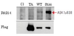
WB图像

( – )泳道:对照质粒

TA泳道:过度表达磷酸化抗体识别部位苏氨酸838使Ala丙氨酸变异的人ASK1。

WT泳道:过度表达野生型人ASK1

Stim泳道:过度表达野生型人ASK1,H2O2刺激激活。

抗磷酸化ASK1,单克隆抗体

数据提供:东京大学大学院药学研究科 丸山顺一 野口拓也

抗原信息

第838苏氨酸磷酸化的人ASK 1 835   – 845氨基酸序列肽

物种交叉反应性

人,小鼠

标签

非标签

抗原别名

Apoptosis Signal Regulating Kinase 1
  ※ASK1的别名
  MAP3K5, MEKK5, MAPKKK5

免疫动物

小鼠

克隆号

PA214

详细信息

ASK1是位于MAP激酶通路最上游的细胞内蛋白质磷酸酶。通过应激刺激使活性酶细胞被激活,第838苏氨酸磷酸化而被激活。诱导凋亡和细胞分化。另一方面有报道称,ASK1活化性可诱导阿尔茨海默症和ALS神经细胞凋亡。

本产品是识别第838苏氨酸磷酸化ASK 1的单克隆抗体。

使用文献

1. Ichijo, H., Nishida, E., Irie, K., et   al.: SCIENCE, 275, 90 (1997).


相关资料


抗磷酸化ASK1,单克隆抗体

抗磷酸化ASK1,单克隆抗体017-22351英文说明书.pdf

(欲了解内容请点击图片)

产品编号 产品名称 产品规格 产品等级 产品价格
017-22351 Anti Phosphorylated ASK1, Monoclonal   Antibody 
抗磷酸化ASK1,单克隆抗体
​50μg 免疫化学用

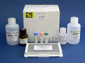
Shibayagi 胰高血糖素样肽-1(GLP-1)(活性) ELISA试剂盒

Shibayagi 胰高血糖素样肽-1(GLP-1)(活性) ELISA试剂盒
Lbis GLP-1(active) ELISA KIT

  • 产品特性
  • 相关资料
  • Q&A
  • 参考文献

Lbis GLP-1(active) ELISA KITShibayagi 胰高血糖素样肽-1(GLP-1)(活性) ELISA试剂盒

Shibayagi 胰高血糖素样肽-1(GLP-1)(活性) ELISA试剂盒

Shibayagi 胰高血糖素样肽-1(GLP-1)(活性) ELISA试剂盒

胰高血糖素样肽-1(Glucagon-like peptide-1,GLP-1)是胰高血糖素前驱体的一部分。胰高血糖素前驱体于胰脏、小肠下部以及下丘脑中表达。该前驱体的构造中含有与糖代谢有关的各种各样的生理活性物质(胰高血糖素,肠高血糖素,胃泌酸调节素,GLP-1,GLP-2)的氨基酸序列。根据表达部位加工酶的特异性,胰脏主要分泌胰高血糖素,而小肠下部主要分泌肠高血糖素和胃泌酸调节素。GLP-1和GLP-2则存在于胰高血糖素前驱体后半的结构中。GLP-1由37个氨基酸组成,有2种生物活性形式,分别为GLP-1(7-37)和GLP-1 (7-36)酰胺。两者都存在于小肠下部、胰脏和下丘脑中,GLP-1(7-36)酰胺在下丘脑中占免疫反应GLP-1(IR-GLP-1)总量的55-94%,在小肠中占27-73%。但在胰脏中只有极少量存在。大部分哺乳类(如人类、大鼠、小鼠、牛、猪、狗等)的GLP-1结构相似。


GLP-1: hdeferhaegtftsdvssylegqaakefiawlvkgrg  

GLP 1(7-37): haegtftsdvssylegqaakefiawlvkgrg

GLP 1(7-36) amide: haegtftsdvssylegqaakefiawlvkgr-NH2


GLP-1与小肠上部分泌的GIP统称为肠促胰素。该类激素是葡萄糖浓度依赖性方式促进胰岛素分泌。同时具有抑制胃肠道蠕动和胃液分泌、抑制胰高血糖素的释放、促进生长抑素的分泌、使食欲减退,促进肠上皮细胞生长、以及外周组织促进非胰岛素依赖性的葡萄糖的消耗,并促进细胞的生长的作用。有报告指出该类激素与垂体激素的分泌也有关系。

GLP-1(7-36)酰胺在生物体内代谢迅速,DPP-IV (dipeptidyl peptidase IV )会使其失去N-末端的两个氨基酸变为GLP-1(9-36)酰胺,GLP-1(7-37)变为GLP-1(9-37)后会失去活性。有报告指出,体外实验中,在犬的血浆中GLP-1(7-36)酰胺的半衰期是为61±9分,GLP-1(7-37)为132±16分。因此GLP-1的测定,取样的时候有必要使用DPP-IV抑制剂。

此外,肠促胰素中的GIP则是最有力促进GLP-1分泌的激素。回肠中GLP-1的分泌不是食物直接刺激肠道而产生的,而是由于胆碱能和肽类的刺激所产生的。

◆特点

● 短时间测定(完全反应时间:5小时)

● 微量样品(标准操作用量:10 μl)可测

● 使用对环境无害的防腐剂

● 全部试剂均为液体,可直接使用

● 精密的测定精度和高再现性

◆构成


组成

状态

容量

(A) 抗体固相化 96 孔板

洗净后使用

96   wells(8×12)/1 块

(B) GLP-1标准溶液(500 pg/mL

稀释后使用

200 μL/1 瓶

(C) 缓冲液

即用

60 mL/1 瓶

(D)生物素结合抗GLP-1抗体

稀释后使用

100 μL/1 瓶

(E) 过氧化物・抗生物素蛋白结合物

稀释后使用

100 μL/1 瓶

(F) 显色液(TMB)

即用

12 mL/1 瓶

(H) 反应终止液(1M H2SO4)※小心轻放

即用

12 mL/1 瓶

( I ) 浓缩洗净液(10×)

稀释后使用

100 mL/1 瓶

封板膜

4 张

使用说明书

1 份

◆交叉反应

※交差率是1,000 pg/mL浓度时的数据

动物类型

对象物质

反应性和反应率(%)

Mouse/Rat

GLP-1(7-36)amide

100

GLP-1(7-37)

<   0.1

GLP-1(1-37)

GLP-1(9-36)amide

GLP-2

Glucagon(1-29)

Insulin

Secretin

GIP

VIP

GRF

Bovine

Glucagon(1-29)

VIP

Porcine

Glucagon(1-29)

VIP

―:不存在交叉反应

◆样品信息


小鼠和大鼠的血清及血浆

10 μL/well(标准操作方法)

※测量中由于酶(DPP-IV等)的影响,采血时请注意防止GLP-1(7-36)酰胺的分解,再使用。


◆测定范围


1.56~50.0 pg/mL 【0.47~15.16 pmol/L(分子量3298)】(标准曲线范围)

7.8~250 pg/mL(样品量10 μ)

3.9~125 pg/mL(样品量20 μ)

◆Validation data


精度测试(检测内变异系数)


样品

A

B

1

23.7

6.44

2

23.2

5.97

3

23.4

6.39

4

24.0

5.87

5

24.1

6.44

mean

23.7

6.22

SD

0.35

0.28

CV(%)

1.5

4.5

单位:pg/mL


再现性测试(检测内变异系数)


测量日/样品

E

F

第0天

25.1

6.31

第1天

25.1

6.16

第2天

25.0

6.24

第3天

25.0

6.37

mean

25.0

6.27

SD

0.03

0.09

CV(%)

0.13

1.4

单位:pg/mL n=4


添加回收测试


样品C

添加量

实测值

回收量

回收率(%)

0.00

3.93

3.26

7.28

3.35

103

6.51

10.3

6.37

97.8

8.14

12.1

8.17

100


样品D

添加量

实测值

回收量

回收率(%)

0.00

11.8

7.16

19.1

7.30

102

14.3

25.5

13.7

95.8

21.5

32.4

20.6

95.8


稀释直线性测试


用稀释缓冲液分三次连续稀释2个血清样品的结果,直线回归方程的R2在0.997~0.9999之间。

相关资料


Shibayagi 胰高血糖素样肽-1(GLP-1)(活性) ELISA试剂盒 Shibayagi 胰高血糖素样肽-1(GLP-1)(活性) ELISA试剂盒

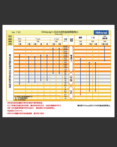
ELISA试剂盒选择指南①②

ELISA试剂盒选择指③④

参考文献

1.

Elevated hepatic DPP4 activity promotes insulin resistance and non-alcoholic fatty liver disease. Baumeier C, Schluter L, Saussenthaler S, Laeger T, Rodiger M, Alaze SA, Fritsche L, Haring HU, Stefan N, Fritsche A, Schwenk RW, Schurmann A. Mol Metab. 2017 Oct;6(10):1254-1263.


 2.

Recombinant Mouse Osteocalcin Secreted by Lactococcus lactis Promotes Glucagon-Like Peptide-1 Induction in STC-1 Cells. Namai F, Shigemori S, Sudo K, Sato T, Yamamoto Y, Nigar S, Ogita T, Shimosato T. Curr Microbiol. 2017 Sep 13.


 3.

A proliferative probiotic Bifidobacterium strain in the gut ameliorates progression of metabolic disorders via microbiota modulation and acetate elevation. Aoki R, Kamikado K, Suda W, Takii H, Mikami Y, Suganuma N, Hattori M, Koga Y. Sci Rep. 2017 Mar 2;7:43522.


 4.

Effect of miglitol on the suppression of nonalcoholic steatohepatitis development and improvement of the gut environment in a rodent model. Kishida Y, Okubo H, Ohno H, Oki K, Yoneda M. J Gastroenterol. 2017 Mar 27.


 5.

Fermented vegetable and fruit extract (OM-XŪ) stimulates murine gastrointestinal tract cells and RAW264. 7 cells in vitro and regulates liver gene expression in vivo. Wakame K, Nakata A, Sato K, Mihara Y, Takahata M, Miyake Y, Okada M, Shimomiya Y, and Komatsu K. Integr Mol Med, 2017 http://www.omx.co.jp/files/attachments/8e3f7824b69bece18acca14baf314b11.PDF

 6.

Gut commensal Bacteroides acidifaciens prevents obesity and improves insulin sensitivity in mice. Yang JY, Lee YS, Kim Y, Lee SH, Ryu S, Fukuda S, Hase K, Yang CS, Lim HS, Kim MS, Kim HM, Ahn SH, Kwon BE, Ko HJ, Kweon MN. Mucosal Immunol. 2017 Jan;10(1):104-116


 7.

An extract from pork bones containing osteocalcin improves glucose metabolism in mice by oral administration. Mizokami A, Wang D, Tanaka M, Gao J, Takeuchi H, Matsui T, Hirata M. Biosci Biotechnol Biochem. 2016 Jul 27:1-8.


 8.

Catecholamines Facilitate Fuel Expenditure and Protect Against Obesity via a Novel Network of the Gut-Brain Axis in Transcription Factor Skn-1-deficient Mice. Ushiama S, Ishimaru Y, Narukawa M, Yoshioka M, Kozuka C, Watanabe N, Tsunoda M, Osakabe N, Asakura T, Masuzaki H, Abe K. EBioMedicine. 2016 Jun;8:60-71.


 9.

Dipeptidyl peptidase 4 inhibitor reduces intimal hyperplasia in rabbit autologous jugular vein graft under poor distal runoff. Koyama A, Komori K, Otsuka R, Kajikuri J, Itoh T. J Vasc Surg. 2016 May;63(5):1360-70.


10.

Dipeptidyl peptidase-4 inhibitor, linagliptin, ameliorates endothelial dysfunction and atherogenesis in normoglycemic apolipoprotein-E deficient mice. Salim HM, Fukuda D, Higashikuni Y, Tanaka K, Hirata Y, Yagi S, Soeki T, Shimabukuro M, Sata M. Vascul Pharmacol. 2016 Apr;79:16-23.


11.

Intestinal Bile Acid Composition Modulates Prohormone Convertase 1/3 (PC1/3) Expression and Consequent GLP-1 Production in Male Mice. Morimoto K, Watanabe M, Sugizaki T, Irie J, Itoh H. Endocrinology. 2016 Mar;157(3):1071-81.


12.

Total gastrectomy-induced reductions in food intake and weight are counteracted by rikkunshito by attenuating glucagon-like peptide-1 elevation in rats. Taguchi M, Dezaki K, Koizumi M, Kurashina K, Hosoya Y, Lefor AK, Sata N, Yada T. Surgery. 2016 Jan 13. pii: S0039-6060(15)01029-6


13.

DPP-4 inhibition has beneficial effects on the heart after myocardial infarction. Akihiko Kubota, Hiroyuki Takano, Haixiu Wang, Hiroshi Hasegawa, Hiroyuki Tadokoro, Masanori Hirose, Yuka Kobaraa, Tomoko Yamada-Inagawa, Issei Komuro, Yoshio Kobayashi. Journal of Molecular and Cellular Cardiology, Volume 91, Feb. 2016, Pages 72–80


14.

The dipeptidyl peptidase IV inhibitor vildagliptin suppresses development of neuropathy in diabetic rodents: Effects on peripheral sensory nerve function, structure and molecular changes. Tsuboi K, Mizukami H, Inaba W, Baba M, Yagihashi S. J Neurochem. Volume 136, Issue 4, pages 859–870, Feb. 2016


15.

Intestinal Bile Acid Composition Modulates Prohormone Convertase 1/3 (PC1/3) Expression and Consequent GLP-1 Production in male mice. Morimoto K, Watanabe M, Sugizaki T, Irie JI, Itoh H. Endocrinology. 2016 Jan 20:en20151551.


16.

Hypoxia decreases glucagon-like peptide-1 secretion from the GLUTag cell line. Kihira Y, Burentogtokh A, Itoh M, Izawa-Ishizawa Y, Ishizawa K, Ikeda Y, Tsuchiya K, Tamaki T. Biol Pharm Bull. Vol.38(4), p514-21, 2015.


17.

Dietary obacunone supplementation stimulates muscle hypertrophy, and suppresses hyperglycemia and obesity through the TGR5 and PPARγ pathway. Horiba T, Katsukawa M, Mita M, Sato R. Biochem Biophys Res Commun. Vol.463(4), p846-52, Aug 2015.


18.

Combination of DPP-4 inhibitor and PPARγ agonist exerts protective effects on pancreatic β-cells in diabetic db/db mice through the augmentation of IRS-2 expression. Hirukawa H, Kaneto H, Shimoda M, Kimura T, Okauchi S, Obata A, Kohara K, Hamamoto S, Tawaramoto K, Hashiramoto M, Kaku K. Mol Cell Endocrinol. Jun 2015.


19.

Glucagon-like peptide-1 is specifically involved in sweet taste transmission. Takai S, Yasumatsu K, Inoue M, Iwata S, Yoshida R, Shigemura N, Yanagawa Y, Drucker DJ, Margolskee RF, Ninomiya Y. FASEB J. Vol.29(6), p2268-80, Jun 2015.


20.

Dipeptidyl-peptidase-4 inhibitor, alogliptin, attenuates arterial inflammation and neointimal formation after injury in low-density lipoprotein (LDL) receptor-deficient mice. Akita K, Isoda K, Shimada K, Daida H. J Am Heart Assoc. Vol.13;4(3):e001469, Mar 2015.


21.

Effects of sleeve gastrectomy and gastric banding on the hypothalamic feeding center in an obese rat model. Kawasaki T1, Ohta M, Kawano Y, Masuda T, Gotoh K, Inomata M, Kitano S.  Surg Today. 2015 Feb 28.


22.

Duodenal-jejunal bypass improves diabetes and liver steatosis via enhanced glucagon-like peptide-1 elicited by bile acids. Kashihara H, Shimada M, Kurita N, Sato H, Yoshikawa K, Higashijima J, Chikakiyo M, Nishi M, Takasu C. J Gastroenterol Hepatol. Vol.30(2), p308-15, Feb 2015.


23.

Mosapride citrate improves nonalcoholic steatohepatitis with increased fecal lactic acid bacteria and plasma glucagon-like peptide-1 level in a rodent model. Okubo H, Nakatsu Y, Sakoda H, Kushiyama A, Fujishiro M, Fukushima T1, Matsunaga Y, Ohno H, Yoneda M, Kamata H, Shinjo T, Iwashita M, Nishimura F, Asano T. Am J Physiol Gastrointest Liver Physiol. Vol.15;308(2), G151-8, Jan 2015.


24.

Glucagon-like peptide-1 production in the GLUTag cell line is impaired by free fatty acids via endoplasmic reticulum stress. Hayashi H, Yamada R, Das SS, Sato T, Takahashi A, Hiratsuka M, Hirasawa N. Metabolism. Vol.63(6), p800-11. Jun 2014.


25.

MK-0626, a selective DPP-4 inhibitor, attenuates hepatic steatosis in ob/ob mice. Ohyama T, Sato K, Yamazaki Y, Hashizume H, Horiguchi N, Kakizaki S, Mori M, Kusano M, Yamada M. World J Gastroenterol.Vol.20(43), p16227-35, Nov 2014.


26.

Oral administration of osteocalcin improves glucose utilization by stimulating glucagon-like peptide-1 secretion. Mizokami A, Yasutake Y, Higashi S, Kawakubo-Yasukochi T, Chishaki S, Takahashi I, Takeuchi H, Hirata M. Bone. 16;69C:68-79. Sep 2014.


27.

Dietary sweet potato (Ipomoea batatas L.) leaf extract attenuates hyperglycaemia by enhancing the secretion of glucagon-like peptide-1 (GLP-1) . Nagamine R, Ueno S, Tsubata M, Yamaguchi K, Takagaki K, Hira T, Hara H, Tsuda T. Food Funct, Vol.5(9), p2309-2316, Aug 2014.


28.

Duodenal-jejunal bypass improves diabetes and liver steatosis via enhanced glucagon-like peptide-1 elicited by bile acids. Kashihara H, Shimada M, Kurita N, Sato H, Yoshikawa K, Higashijima J, Chikakiyo M, Nishi M, Takasu C. Journal of Gastroenterology and Hepatology, Aug 2014.


29.

Deletion of Hypoxia-Inducible Factor-1α in Adipocytes Enhances Glucagon-Like Peptide-1 Secretion and Reduces Adipose Tissue Inflammation. Kihira Y, Miyake M, Hirata M, Hoshina Y, Kato K, Shirakawa H, Sakaue H, Yamano N, Izawa-Ishizawa Y, Ishizawa K, Ikeda Y, Tsuchiya K, Tamaki T, Tomita S. PLoS One. 2014 Apr 4;9(4):e93856


30.

Glucagon-like peptide-1 production in the GLUTag cell line is impaired by free fatty acids via endoplasmic reticulum stress. Hayashi H., Yamada R., Shankar Das S., Sato T., Takahashi A., Hiratsuka M., Hirasawa N. Metabolism – Clinical and Experimental, 2014.


31.

Dipeptidyl peptidase-4 inhibitor ameliorates early renal injury through its anti-inflammatory action in a rat model of type 1 diabetes. Kodera AR, Shikata K., Takatsuka T., Oda K., Miyamoto S., Kajitani N., Hirota D., Ono T., Usui HK., Makino H. Biochemical and Biophysical Research Communications, Vol.443(3), p828-833, Jan 2014.


32.

Novel insight into the distribution of L-cells in the rat intestinal tract. Hansen CF., Vrang N., Sangild PT., Jelsing J. Am J Transl Res, Vol.5(3), p347-358, 2013.


33.

Cinnamtannin A2, a Tetrameric Procyanidin, Increases GLP-1 and Insulin Secretion in Mice. Yamashita Y, Okabe M, Natsume M, Ashida H. Bioscience, Biotechnology, and Biochemistry , Vol.77(4), 2013


34.

The protective roles of GLP-1R signaling in diabetic nephropathy:possible mechanism and therapeutic potential. H.Fujita, T.Morii, H.Fujishima, T.Sato, T.Shimizu, M.Hosoba, K.Tsukiyama, T.Narita, T.Takahashi, D.J.Drucker, Y.Seino, and Y.Yamada. Kidney International, 2013


35.

Beneficial effects of vildagliptin combined with miglitol on glucose tolerance and islet morphology in diet-controlled db/db mice. K.Ishibashi., A.Hara., Y.Fujitani., T.Uchida., K.Komiya., M.Tamaki., H.Abe., T.Ogihara., A.Kanazawa., R.Kawamori and H.Watada. Biochem Biophys Res Commun, Vol.440(4), p570-575, Nov 2013.


36.

Vildagliptin preserves the mass and function of pancreatic β cells via the developmental regulation and suppression of oxidative and endoplasmic reticulum stress in a mouse model of diabetes. Hamamoto S, Kanda Y, Shimoda M, Tatsumi F, Kohara K, Tawaramoto K, Hashiramoto M and Kaku K. Diabetes, Obesity and Metabolism,Vol.15(2), p153-163, Feb 2013.


37.

Osteocalcin Induces Release of Glucagon-Like Peptide-1 and Thereby Stimulates Insulin Secretion in Mice. Mizokami A, Yasutake Y, Gao J, Matsuda M, Takahashi I, Takeuchi H and Hirata M. PLoS ONE 8(2): e57375. Feb 2013


38.

DPP4 inhibitor vildagliptin preserves β-cell mass through amelioration of endoplasmic reticulum stress in C/EBPB transgenic mice. Shinobu Shimizu, Tetsuya Hosooka, Tomokazu Matsuda, Shun-ichiro Asahara, Maki Koyanagi-Kimura, Ayumi Kanno, Alberto Bartolome, Hiroaki Etoh, Megumi Fuchita, Kyoko Teruyama, Hiroaki Takahashi, Hiroyuki Inoue, Yusuke Mieda, Naoko Hashimoto, Susumu Seino, and Yoshiaki Kido. J Mol Endocrinol, Vol.49, p125-135, Oct 2012.


39.

Effects of long-term treatment with the dipeptidyl peptidase-4 inhibitor vildagliptin on islet endocrine cells in non-obese type 2 diabetic Goto-Kakizaki rats. Inaba W, Mizukami H, Kamata K, Takahashi K, Tsuboi K and Yagihashi S. European Journal of Pharmacology,Vol.691(1-3), p297-306, Sep 2012.


40.

The DPP4 inhibitor linagliptin delays the onset of diabetes and preserves β-cell mass in non-obese diabetic mice. Jacob Jelsing, Niels Vrang, Soren B van Witteloostuijn, Michael Mark and Thomas Klein. J Endocrinol, Vol.214, p381-387, Sep 2012.


41.

Neural and humoral changes associated with the adjustable gastric band: insights from a rodent model. J Kampe, A Stefanidis, S H Lockie, W A Brown, J B Dixon, A Odoi, S J Spencer, J Raven and B J Oldfield. International Journal of Obesity, 27 Mar 2012.


42.

Neural and humoral changes associated with the adjustable gastric band: insights from a rodent model. J Kampe, A Stefanidis, S H Lockie, W A Brown, J B Dixon, A Odoi, S J Spencer, J Raven and B J Oldfield. International Journal of Obesity, 27 Mar 2012.


43.

Mate Tea(Ilex paraguariensis)Promotes Satiety and Body Weight Lowering in Mice:Involvement of Glucagon-Like Peptide-1. G, M, E, Hussein., H, Matsuda., S, Nakamura., M, Hamao., T, Akiyama., K, Tamura., and M, Yoshikawa. Biol.Pharm.Bull. Vol.34(12), p1849-1855, 2011.


44.

5-Hydroxy-eicosapentaenoic acid is an endogenous GPR119 agonist and enhances glucose-dependent insulin secretion. R, Kogure., K, Toyama., S, Hiyamuta., I, Kojima., S, Takeda.  Biochemical and Biophysical Research Communications Vol.416(1-2), p58-63, 2011.


45.

GLP-1 Secretion in Response to Oral and Luminal Palatinose (Isomaltulose) in Rats. T,Hira.,M,Muramatsu.,M,Okuno.and H,Hara. J Nutr Sci Vitaminol, Vol.57, p30-35, 2011.


46.

Trehalose prevents adipocyte hypertrophy and mitigates insulin resistance. C,Arai.,N,Arai.,A,Mizote.,K,Kohno.,K,Iwaki.,T,Hanaya.,S,Arai.,S,Ushio.,S,Fukuda. Nutrition Research, Vol.30(12), p840-848, 2010.


47.

Imaging exocytosis of single glucagon-like peptide-1 containing granules in a murine enteroendocrine cell line with total internal reflection fluorescent microscopy. Ohara-Imaizumi,M.,Aoyagi,K.,Akimoto,Y.,Nakamichi,Y.,Nishiwaki,C.,Kawakami,H.and Nagamatsu,S. Biochemical and Biophysical Reseach Communications, Vol.390, p16-20, 2009.


产品编号 产品名称 产品规格 产品等级 产品价格
637-15129 (AKMGP-011) Lbis® GLP-1(Active) ELISA Kit
Lbis® 胰高血糖素样肽-1(GLP-1)(活性) ELISA试剂盒 
96 tests

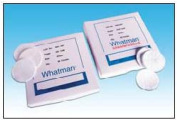
1823-150-Whatman 玻璃微纤维滤纸 Grade GF/D 150mm-玻璃纤维滤膜

品牌 其他品牌 货号 1823-150
供货周期 现货 应用领域 医疗/卫生,环保/水工业,生物产业,综合

Whatman 玻璃微纤维滤纸 Grade GF/D 150mm
流速相当快,在相同颗粒保留度的条件下,整个过滤速度比纤维素更快。滤纸较厚,具有较强的负载力,可作为膜的预滤。由不同大小来适合大多数滤器。GF/D为保留细小颗粒的膜提供了很好的保护,可以和GF/B一起使用,给膜提供了非常有效的预滤保护。

Whatman 玻璃微纤维滤纸 Grade GF/D 150mm

Grade GF/D:2.7µm

流速相当快,在相同颗粒保留度的条件下,整个过滤速度比纤维素更快。滤纸较厚,具有较强的负载力,可作为膜的预滤。由不同大小来适合大多数滤器。GF/D为保留细小颗粒的膜提供了很好的保护,可以和GF/B一起使用,给膜提供了非常有效的预滤保护。

Whatman 玻璃微纤维滤纸 Grade GF/D 150mm技术参数

整包数量:    100 片

等级:    Grade GF/D1)

直径:    150mm

液体中的颗粒保留:    2.7 µm

过滤速度:    快速

气体流速:    2.2s/100ml/in2

常规厚度:    675μm

基本重量:    121g/m2

材质:    硼硅酸玻璃

属性特点:    高负载能力

黏合剂类型:    无黏合剂

订购信息:

1823-025 GF/D 2.5CM 100/PK
1823-042 GF/D 4.25CM 100/PK
1823-047 GF/D 4.7CM 100/PK
1823-055 GF/D 5.5CM 100/PK
1823-070 GF/D 7CM 100/PK
1823-090 GF/D 9CM 25/PK
1823-110 GF/D 11CM 25/PK
1823-125 GF/D 12.5CM 25/PK
1823-142 GF/D 14.2CM 25/PK
1823-150 GF/D 15CM 25/PK
1823-257 GF/D 25.7CM 25/PK

 是国内专业的实验过滤材料提供商,致力于将质量、可靠性和操作性突出的产品带给每一位客户。
公司产品包括各类滤纸、滤膜、滤器以及辅助器材,主营定性滤纸,定量滤纸,层析纸,玻璃微纤维滤纸;石英滤膜,无机膜,聚碳酸酯膜(PC),聚四氟乙烯(PTFE)滤膜,聚偏二氟乙烯(PVDF)膜,尼龙(Nylon)膜,聚醚砜(PES)膜,醋酸纤维素(CA)膜,硝酸纤维素(NC)膜,混合纤维素膜(MCE);针头式滤器,囊式滤器,可换膜过滤器,超滤离心管等。广泛应用于生命科学,农业,药物医学,化工,食品,水质分析,环境监测等各个领域,公司与各全球知名厂家建立了稳定的合作关系,确保正品的同时更可以满足客户对于便捷和实惠的需求。

伯乐iScript Select cDNA合成试剂盒1708897

Bio-Rad伯乐iScript Select cDNA合成试剂盒1708897

iScript Select cDNA 合成试剂盒是一种灵敏的逆转录系统,提供灵活的引发策略。与我们的 iScript cDNA 合成试剂盒和用于 RT-qPCR 的 iScript 逆转录试剂盒不同,此试剂盒带有多种反应组分(5 倍缓冲液,低聚糖 [dT]、随机引物、专有基因特定的引物强化剂溶液和水),必须在反应设置过程中合并。

主要功能和优点

  • mRNA 灌注策略具有灵活性 — 用单独的试管提供低聚糖 (dT)、随机引物和基因特定的引物强化剂
  • 合成最大 8 kb 的长 cDNA — 通过质量控制将 cDNA 的可靠合成长度控制为 >6 kb
  • 检测低水平靶基因并在基因表达分析过程中保存 RNA — 广泛的线性动态范围内的总输入 RNA (1 µg–1 pg),带有高效的 RNase H+ MMLV 逆转录酶
  • 避免从 RNA 降解获得有偏颇的数据 — RNaseA 抑制剂的强效混合物可在设置和逆转录过程中保护 RNA
  • 恒温冷冻室中在 –20°C 下可以保存 12 个月(不建议多次解冻)

1708889:iScript cDNA 合成反应试剂,5x,100个反应

1708890:iScript cDNA 合成试剂盒,25x20ul

1708891:iScript cDNA 合成试剂盒,100x20ul

1708896:iScript 选择型cDNA 合成试剂盒,25x20ul

1708897:iScript 选择型cDNA 合成试剂盒,100x20ul

Bio-Rad伯乐iScript Select cDNA合成试剂盒1708897


fisherbrand带缺口塑料烧杯 190ml CONTAINR FIS14-955-112-赛默飞中国代理商

产品信息
产品名称:
fisherbrand带缺口塑料烧杯 190ml CONTAINR FIS14-955-112
产品型号:
fisherbrand带缺口塑料烧杯 190ml CONTAINR FIS14-955-112 产品特点
  美国Fisherbrand带缺口烧杯 适用于盛装尿液或痰 ● 透明聚苯乙烯材质,带倾倒口 ● 适用于采集少量液体样本及试纸检测 ● 可耐受稀酸或弱酸 ● 可叠放,带 oz. 、cc 和 mL 刻度

fisherbrand带缺口塑料烧杯 190ml CONTAINR FIS14-955-112
产品详细信息:

美国Fisherbrand带缺口烧杯 CONTAINR 6.5OZ W/O LIDS 500/CS

适用于盛装尿液或痰
● 透明聚苯乙烯材质,带倾倒口
● 适用于采集少量液体样本及试纸检测
● 可耐受稀酸或弱酸
● 可叠放,带 oz. 、cc 和 mL 刻度

产品型号:FIS#14-955-112 190ml 500个/箱

  • fisherbrand带缺口烧杯
  • CONTAINR 6.5OZ W/O LIDS 500/CS带倾倒口烧杯,透明聚苯乙烯材质,6.5 oz., 500个/箱
  • 单位:箱(500个/箱)
  • 预计45-60Days
  • ¥666.0元
  • ¥555.0元
  • 1
  •  
  • 加入
  • fisherbrand带缺口烧杯盖子
  • LID FOR 14-955-112 25/PK烧杯盖子, 25个/盒,500个/箱
  • 单位:箱(500个/箱)
  • 预计45-60Days
  • ¥422.0元
  • ¥311.0元
  • 1
  •  
  • 加入

小胶质细胞/巨噬细胞特异性蛋白抗体–Iba1抗体,兔(免疫组化)

小胶质细胞/巨噬细胞特异性蛋白抗体–Iba1抗体,兔(免疫组化)
Anti Iba1, Rabbit (for Immunocytochemistry)

  • 产品特性
  • 相关资料
  • Q&A
  • 参考文献

小胶质细胞/巨噬细胞特异性蛋白抗体–Iba1抗体,兔(免疫组化)小胶质细胞/巨噬细胞特异性蛋白抗体--Iba1抗体,兔(免疫组化)

Anti Iba1, Rabbit (for   Immunocytochemistry)


小胶质细胞标记抗体(免疫染色用)

产品编号

产品名称【中文名称】

规格

包装

019-19741

Anti Iba1, Rabbit (for   Immunocytochemistry)

【抗Iba1,兔(免疫细胞化学)】

免疫化学用

50 μg

抗体信息

抗原名

Iba1

适用

实验

免疫

染色

同种型

IgG

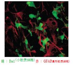
免疫染色图像

小胶质细胞/巨噬细胞特异性蛋白抗体--Iba1抗体,兔(免疫组化)

抗原信息

Iba1C末端序列肽

物种交叉反应性

人,小鼠,大鼠

标签

非标签

抗原别名

AIF-1, IRT1, Protein G1

免疫

动物

克隆号


(多克隆抗体)

详细信息

Iba1是与巨噬细胞/小胶质细胞特异性表达,分子量17000的钙结合蛋白。近年来,小胶质细胞除了对神经营养·保护作用以外,产生的NO、TNF- α、IL-1 β对神经伤害作用很受关注。

本产品是与小胶质细胞特异性反应的兔多克隆抗体,适用于与星形胶质细胞特异性GFAP单克隆抗体的双重染色。

使用文献

[1] Jun,et al.(2017).Nature,551(7679),232-236.

[2] Gibson,et al.(2014).Science,344(6183),1252304.

[3] Yan,et al. (2018).Cell, S009286741830285X.

[4] Pil,et al. (2018). Nature Medicine.

欲了解相关资料请点击文字:

Wako神经生物学抗体清单

巨噬细胞/小胶质细胞Iba1抗体

◆相关资料


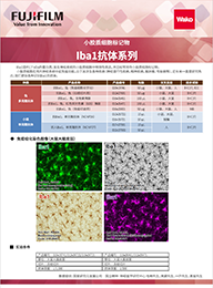
Iba1抗体选择指南


小胶质细胞/巨噬细胞特异性蛋白抗体--Iba1抗体,兔(免疫组化)

小胶质细胞/巨噬细胞特异性蛋白抗体--Iba1抗体,兔(免疫组化)Iba1抗体选择指南.pdf

参考文献


◆Nature


 1.

Lam, C.K., et al.: Nature, 465, 478(2010).

Embolus extravasation is an alternative mechanism for cerebral microvascular recanalization.

 2.

Stefater, J. A. 3rd. et al.: Nature, 474, 511(2011).   

Regulation of angiogenesis by a non-canonical Wnt-Flt1 pathway in myeloid cells

 3.

Deng, H. X., et al.: Nature, 477, 211(2011).

Mutations in UBQLN2 cause dominant X-linked juvenile and adult onset ALS and ALS/dementia.

 4.

Lee, Y., et al.: Nature, 487, 433(2012).

Oligodendroglia metabolically support axons and contribute to neurodegeneration.

 5.

Heneka, M. T., et al.: Nature, 493, 674(2013).

NLRP3 is activated in Alzheimer's disease and contributes to pathology in APP/PS1 mice.

 6.

Shao, W., et al.: Nature, 494, 90(2013).

Suppression of neuroinflammation by astrocytic dopamine D2 receptors via αB-crystallin

 7.

Zhang, G., et al.: Nature, 497, 211(2013).

Hypothalamic programming of systemic ageing involving IKK-β, NF-κB and GnRH

 8.

Chung, W. S., et al.: Nature, 504, 394(2013).

Astrocytes mediate synapse elimination through MEGF10 and MERTK pathways.

 9.

Roth, T. L., et al.: Nature, 505, 223(2014).

Transcranial amelioration of inflammation and cell death after brain injury

10.

Najm, F. J., et al.: Nature, 522, 216(2015).

Drug-based modulation of endogenous stem cells promotes functional remyelination in vivo.


11.

Fourgeaud, L., et al.: Nature, 532, 240(2016).

TAM receptors regulate multiple features of microglial physiology.

12.

Vasek, M. J., et al.: Nature, 534, 538(2016).

A complement-microglial axis drives synapse loss during virus-induced memory impairment.


13.

Iaccarino, H. F., et al.: Nature, 540, 230(2016).

Gamma frequency entrainment attenuates amyloid load and modifies microglia.

14.

Bialas, A. R. et al.: Nature, 546, 539(2017).

Microglia-dependent synapse loss in type I interferon-mediated lupus

15.

Mass, E., et al.: Nature, 549, 389(2017).

A somatic mutation in erythro-myeloid progenitors causes neurodegenerative disease.

16.

Jun, J. J., et al.: Nature, 551, 232(2017).

Fully integrated silicon probes for high-density recording of neural activity.

17.

Bussian, T. J., et al.: Nature, 562, 578(2018).
Clearance of senescent glial cells prevents tau-dependent pathology and cognitive decline.

◆CELL


 1.

Lujambio, A., et al.: Cell, 153, 2, 449(2013).

Non-Cell-Autonomous Tumor Suppression by p53.

 2.

Parkhurst, C. N., et al.: Cell, 155, 7, 1596(2013).

Microglia promote learning-dependent synapse formation through brain-derived neurotrophic

factor.

 3.

Wang, Y., et al.: Cell, 160, 6, 1061(2015).

TREM2 Lipid Sensing Sustains the Microglial Response in an Alzheimer’s Disease Model.

 4.

Keren-Shaul, H., et al.: Cell, 169, 7, 1276(2017).

A Unique Microglia Type Associated with Restricting Development of Alzheimer's Disease.

 5.

Ulland, T. K., et al.: Cell, 170, 4, 649(2017).

TREM2 Maintains Microglial Metabolic Fitness in Alzheimer’s Disease.

 6.

Qin, Y., et al.: Cell, 174, 1, 156(2018).

A Milieu Molecule for TGF-β Required for Microglia Function in the Nervous System.

 7.

Yan, S., et al.: Cell, 173, 4, 989(2018).

A Huntingtin Knockin Pig Model Recapitulates Features of Selective Neurodegeneration in

Huntington's Disease

◆Nature Medicine


 1.

Heppner, F. L., et al.: Nat. Med., 2, 146(2005).  

Experimental autoimmune encephalomyelitis repressed by microglial paralysis.

 2.

Nikić, I., et al.: Nat. Med., 4, 495(2011).

A reversible form of axon damage in experimental autoimmune encephalomyelitis and multiple

sclerosis.


 3.

Vom, B. J., et al.: Nat. Med., 12, 1812(2012).

Inhibition of IL-12/IL-23 signaling reduces Alzheimer’s disease–like pathology and cognitive

decline

 4.

Minami, S. S., et al.: Nat. Med., 10, 1157(2014).

Progranulin protects against amyloid β deposition and toxicity in Alzheimer's disease mouse

models.

 5.

Yun, S. P., et al.: Nat. Med., 7, 931(2018).

Block of A1 astrocyte conversion by microglia is neuroprotective in models of Parkinson's disease.

 6.

Mount, C. W., et al.: Nat Med. 5, 572(2018).

Potent antitumor efficacy of anti-GD2 CAR T cells in H3-K27M+ diffuse midline gliomas.


◆Nature Neuroscience


 1.

Zhang, K., et al.: Nat. Neurosci., 10, 1064(2003).

HIV-induced metalloproteinase processing of the chemokine stromal cell derived factor-1 causes

neurodegeneration.

 2.

Ajami, B., et al.: Nat. Neurosci., 12, 1538(2007).

Local self-renewal can sustain CNS microglia maintenance and function throughout adult life

 3.

Mildner, A., et al.: Nat. Neurosci., 12, 1544(2007).

Microglia in the adult brain arise from Ly-6ChiCCR2+ monocytes only under defined host

conditions.

 4.

Bero, A. W., et al.: Nat. Neurosci., 6, 750(2011).

Neuronal activity regulates the regional vulnerability to amyloid-β deposition.

 5.

Fancy, S. P., et al.: Nat. Neurosci., 14, 1009(2011).

Axin2 as regulatory and therapeutic target in newborn brain injury and remyelination.

 6.

Ajami, B., et al.: Nat. Neurosci., 14, 1142(2011).

Infiltrating monocytes trigger EAE progression, but do not contribute to the resident microglia pool.

 7.

Mosher, K. I. et al.: Nat. Neurosci., 11, 1485(2012).

Neural progenitor cells regulate microglia functions and activity.

 8.

Lehmann, S. M., et al.: Nat. Neurosci., 6, 827(2012).

An unconventional role for miRNA: let-7 activates Toll-like receptor 7 and causes

neurodegeneration.


 9.

Kierdorf, K., et al.: Nat. Neurosci., 3, 273(2013).

Microglia emerge from erythromyeloid precursors via Pu.1- and Irf8-dependent pathways

10.

Bialas, A. R. et al.: Nat. Neurosci., 12, 1773(2013).

TGF-β signaling regulates neuronal C1q expression and developmental synaptic refinement

11.

Butovsky, O., et al.: Nat. Neurosci., 1, 131(2014).

Identification of a Unique TGF-β Dependent Molecular and Functional Signature in Microglia.

12.

Saito, T., et al.: Nat. Neurosci., 5, 661(2014).

Single App knock-in mouse models of Alzheimer's disease.

13.

Erny, D., et al.: Nat. Neurosci., 7, 965(2015).

Host microbiota constantly control maturation and function of microglia in the CNS.


14.

Sorge, R. E. et al.: Nat. Neurosci., 8, 1081(2015).

Different immune cells mediate mechanical pain hypersensitivity in male and female mice.

15.

Hama, H., et al.: Nat. Neurosci., 10, 1518(2015).

ScaleS: an optical clearing palette for biological imaging.

16.

Asai, H., et al.: Nat. Neurosci., 11, 1584(2015).

Depletion of microglia and inhibition of exosome synthesis halt tau propagation.

17.

Guan, Z., et al.: Nat. Neurosci., 1, 94(2016).

Injured sensory neuron-derived CSF1 induces microglial proliferation and DAP12-dependent pain.

18.

Grabert, K., et al.: Nat. Neurosci., 3, 504(2016).

Microglial brain region-dependent diversity and selective regional sensitivities to aging

19.

Gonçalves, J. T., et al.: Nat. Neurosci., 6, 788(2016).

In vivo imaging of dendritic pruning in dentate granule cells

20.

Liu, Q., et al.: Nat. Neurosci., 2, 243(2016).

Neural stem cells sustain natural killer cells that dictate recovery from brain inflammation.

21.

Safaiyan, S., et al.: Nat. Neurosci., 8, 995(2016).

Age-related myelin degradation burdens the clearance function of microglia during aging.

22.

Pandya, H., et al.: Nat. Neurosci., 5, 753(2017).

Differentiation of human and murine induced pluripotent stem cells to microglia-like cells

23.

Füger, P., et al.: Nat. Neurosci., 10, 1371(2017).

Microglia turnover with aging and in an Alzheimer's model via long-term in vivo single-cell imaging


◆Nature Immunology


 1.

Wang, Y., et al.: Nat. Immunol., 13, 753(2012).

IL-34 is a tissue-restricted ligand of CSF1R required for the development of Langerhans cells and

microglia.

 2.

Goldmann, T., et al.: Nat. Immunol., 17, 797(2016).

Origin, fate and dynamics of macrophages at central nervous system interfaces


 3.

Haimon, Z., et al.: Nat. Immunol., 19, 636(2018).

Re-evaluating microglia expression profiles using RiboTag and cell isolation strategies.

◆Nature Biotechnology


 1.

Park, S. I., et al.: Nat. Biotechnol., 33, 1280(2015).

Soft, stretchable, fully implantable miniaturized optoelectronic systems for wireless optogenetics

 2.

Staahl, B. T., et al.: Nat. Biotechnol., 35, 431(2017).

Efficient genome editing in the mouse brain by local delivery of engineered Cas9 ribonucleoprotein

complexes

◆Nature Methods


 1.

Clark, J. J., et al.: Nat. Methods., 7, 126(2010).

Chronic microsensors for longitudinal, subsecond dopamine detection in behaving animals

 2.

Prevedel, R., et al.: Nat. Methods., 13, 1021(2016).
Fast volumetric calcium imaging across multiple cortical layers using sculpted light

◆Neuron


 1.

Simard, A. R., et al.: Neuron, 49, 4, 489(2006).

Bone marrow-derived microglia play a critical role in restricting senile plaque formation in

Alzheimer's disease.

 2.

Bhaskar, K., et al.: Neuron, 68, 1, 19(2010).

Regulation of tau pathology by the microglial fractalkine receptor.

 3.

Bergmann, O., et al.: Neuron, 74, 4, 634(2012).

The Age of Olfactory Bulb Neurons in Humans

 4.

Schafer, D. P., et al.: Neuron, 74, 4, 691(2012).

Microglia sculpt postnatal neural circuits in an activity and complement-dependent manner.

 5.

Paolicelli, R. C., et al.: Neuron, 95, 2, 297(2017).

TDP-43 Depletion in Microglia Promotes Amyloid Clearance but Also Induces Synapse Loss.

 6.

Tufail, Y., et al.: Neuron, 93, 3, 574(2017).

Phosphatidylserine Exposure Controls Viral Innate Immune Responses by Microglia.

 7.

Abud, E. M., et al.: Neuron, 94, 2, 278(2017).

iPSC-Derived Human Microglia-like Cells to Study Neurological Diseases.

 8.

Bohlen, C. J., et al.: Neuron, 94, 4, 759(2017).

Diverse Requirements for Microglial Survival, Specification, and Function Revealed by

Defined-Medium Cultures.

 9.

De, Biase, L. M., et al.: Neuron, 95, 2, 341(2017).

Local Cues Establish and Maintain Region-Specific Phenotypes of Basal Ganglia Microglia.

10.

Hwang, H. W., et al.: Neuron, 95, 6, 1334(2017).

cTag-PAPERCLIP Reveals Alternative Polyadenylation Promotes Cell-Type Specific Protein Diversity

and Shifts Araf Isoforms with Microglia Activation.

11.

Lehrman, E. K., et al.: Neuron, 100, 1, 120(2018).

CD47 Protects Synapses from Excess Microglia-Mediated Pruning during Development.

12.

López-Erauskin, J., et al.: Neuron, 100, 4, 816(2018).

ALS/FTD-Linked Mutation in FUS Suppresses Intra-axonal Protein Synthesis and Drives Disease

Without Nuclear Loss-of-Function of FUS

产品编号 产品名称 产品规格 产品等级 产品价格
019-19741 Anti Iba1, Rabbit (for   Immunocytochemistry) 
抗Iba1,兔(免疫细胞化学)
50μg 免疫化学用

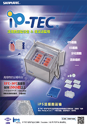
iP-TEC® 36-蓄热板

iP-TEC® 36-蓄热板

  • 产品特性
  • 相关资料
  • Q&A
  • 参考文献

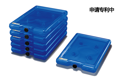
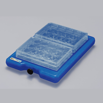
稳定且长时间保持33℃~36℃iP-TEC® 36-蓄热板

可重复使用的托盘型高性能蓄热材料(产品编号:28454)

iP-TEC® 36-蓄热板

  突出的温度维持力,可将温度长时间维持在36℃左右,可反复使用。托盘型的容器形状,不仅适用于定温运输,还可作为保温托盘用于实验室中。


◆温度调节方法(参考)

首先在45℃的环境下将各块蓄热板隔开一定距离,静置4~8小时左右,直至蓄热板内部物质完全溶解为液态状。

然后在36℃的环境下放置1小时左右,或者在室温(20~25℃)下静置10~15分钟,检查表面温度稳定在36℃后使用。

请使用恒温箱干热灭菌器恒温干燥机暖柜等(可设定45℃的设备)进行调温。

※请勿使用架板比设定温度要高的调温设备。


关于测量蓄热板温度方法

推荐使用红外线温度计

◆特点


• 容器材质:PVC   

• 尺寸(mm):230×162×29H   

• 重量:650g

• 可进行酒精喷雾消毒

(注)不能高压灭菌

iP-TEC® 36-蓄热板

iP-TEC® 36-蓄热板

并放2块孔板,可在工作台内边保温边作业。

可稳固堆叠。

◆相关产品

蓄热板专用直立架子(8块用)

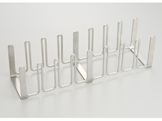
产品编号:WEB28522

iP-TEC® 36-蓄热板

————————————————————————————————————————

蓄热板调温箱HC-INC50(-15℃~50℃)

产品编号:WEB28521

iP-TEC® 36-蓄热板

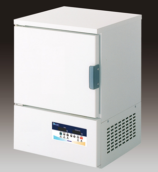
可设定-15℃~50℃,适用于调温各种温度带的蓄热板。

————————————————————————————————————————

蓄热板调温BOX套装(含6块36-蓄热板)

产品编号:WEB28524

iP-TEC® 36-蓄热板

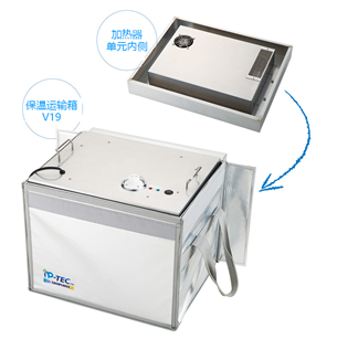
套装内容:1个加热器单元、1个保温运输箱-V19、6块36-蓄热板和1个蓄热板专用直立架子(6块用)。

把加热器单元嵌入保温运输箱-V19中使用。将温度控制旋钮调到“ミドル”,箱内温度自动调节到47℃。36-蓄热板在该环境下放置约7~8小时就能完全溶解。(请参考36-蓄热板溶解后的处理方法。)该套装可一次性对6块蓄热板进行调温,请使用配套的专用架。


iP-TEC® 36-蓄热板 iP-TEC® 36-蓄热板
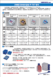
iP-TEC® 活细胞运输系列产品详情 iP-TEC® 系列产品简介列表
iP-TEC® 36-蓄热板 iP-TEC® 36-蓄热板
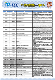
iP-TEC® 问题集 蓄热板安装手册

1、

Q:

是否可以使用微波炉对蓄热板进行调温?
A:
绝对不可以。最好使用恒温箱等恒温设备进行调温。24-蓄热板可用热水进行短时间的调温。
2、

Q:

蓄热板调温有什么技巧吗?
A:
尽可能把蓄热板表面都暴露在恒温环境下,重叠的话,接触面就无法很好地传递温度。技巧是把蓄热板立起来并各自隔开一定距离,留出空间进行调温。
3、

Q:

蓄热板的使用寿命是多久?
A:
一般来说,蓄热板是半永久性可重复使用的。但是,内部物质若混入水分或氧化等会引起性能下降。蓄热板表面破损或可见划痕增多时就应该更换新的产品。
4、

Q:

蓄热板的材料是什么?
A:
属于石蜡材料,详细不便公开。敬请原谅。材料本身符合航空法(日本法规)规定的非危险品,安全可空运。
5、

Q:

蓄热板可以带入飞机客舱吗?
A:
属于非危险品可带入机舱,但对于有温度控制的物品各航空公司规定不一。建议搭乘时咨询航空公司。此外,提供细胞和培养液、蓄热板等各种材料证明时,细胞和培养液需要医师等开具证明其为非危险品。并且,运输国外需要有英文版的上述说明书。建议事前向航空公司或目标客户确认清楚。
6、
Q: 蓄热板可以带入飞机货舱吗?
A:
可以,属于航空法规定的非危险品。但提供细胞和培养液、蓄热板等各种材料证明时,细胞和培养液需要医师等开具证明其为非危险品。并且,运输国外需要有英文版的上述说明书。建议事前向航空公司或目标客户确认清楚。
7、
Q: 蓄热板可以使用水浴调温吗?
A:
可以。但是容器的材质是PVC,水浴温度请不要超过55℃。此外,需根据水浴设备和使用环境调整调温时间。通过观察蓄热板内部物质的溶解程度进行调温工作。

产品编号 产品名称 产品规格 产品等级 产品价格
WEB28454 iP-TEC® 36-蓄热板 1片
WEB28522 蓄热板专用直立架子(8块用) 1个
WEB28521 蓄热板调温箱HC-INC50(-15℃~50℃) 1台
WEB28524 蓄热板调温BOX套装(含6块36-蓄热板) 1套

Whatman05-714-4滤纸、滤器、微孔板系列THK CHR PAPER 46x57CM 100/PK

【简单介绍】

Whatman,实验室高端分离产品的开发和创新者。从滤纸和滤器,到微生物学和色谱产品,Whatman创新的产品和方法使全球生命科学领域中的科学家,工程师和医护工作者能够更简便,更快捷,更精确,更安全的进行他们的工作。无论是在药物成分分析,物质纯化和鉴定,还是环境应用中的非细胞样品制备,无论何处何地,Whatman公司以其高质量,可靠和创新的产品闻名于世。

【简单介绍】

Whatman,实验室高端分离产品的开发和创新者。从滤纸和滤器,到微生物学和色谱产品,Whatman创新的产品和方法使全球生命科学领域中的科学家,工程师和医护工作者能够更简便,更快捷,更精确,更安全的进行他们的工作。无论是在药物成分分析,物质纯化和鉴定,还是环境应用中的非细胞样品制备,无论何处何地,Whatman公司以其高质量,可靠和创新的产品闻名于世。

【详细说明】

原装进口英国Whatman05-714-4滤纸、滤器、微孔板系列THK CHR PAPER 46x57CM 100/PK

英国Whatman05-714-4滤纸、滤器、微孔板系列THK CHR PAPER 46x57CM 100/PK

产品介绍:

主要产品:

l 滤纸和滤膜

l 过滤器具

l 萃取产品

l 层析产品

l 微生物检测产品

l FTA

l 903滤纸

订货信息:

CATALOG NO型号

DESCRIPTION描述

5401-090

Private Label and 2nd brands

464

GF Reeve Angel

401 9CM 100/PK

5401-110

Private Label and 2nd brands

464

GF Reeve Angel

401 11CM 100/PK

5401-125

Private Label and 2nd brands

464

GF Reeve Angel

401 12.5CM 100/PK

5403-047

Private Label and 2nd brands

464

GF Reeve Angel

403 4.7CM 100/PK

5403-090

Private Label and 2nd brands

464

GF Reeve Angel

403 9CM 100/PK

05-714-1

Private Label and 2nd brands

472

CF Private Label

THIN CHR PAPER 46x57CM 100/PK

05-714-4

Private Label and 2nd brands

472

CF Private Label

THK CHR PAPER 46x57CM 100/PK

上海金畔生物科技有限公司

文章号19582198-19582198

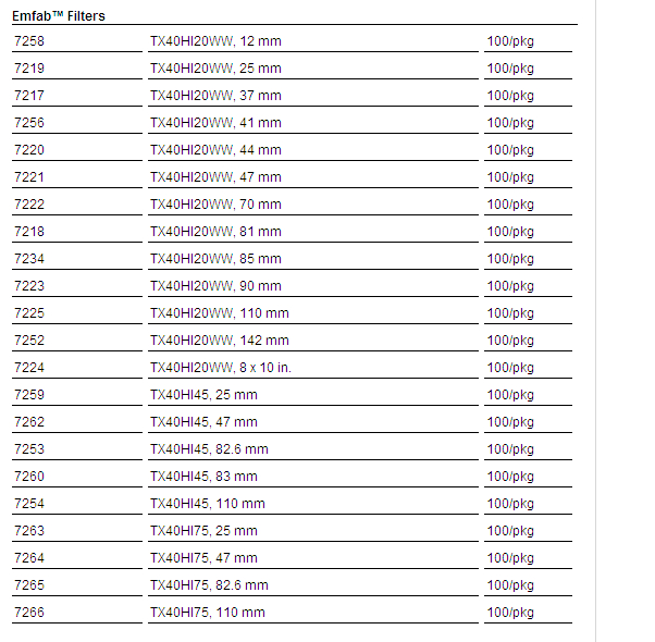
Emfab过滤膜TX40HI20-WW(PALL

Emfab过滤膜TX40HI20-WW(PALL 7221)汽车尾气检测废气检测用过滤膜7221

1、可折叠,有利于称重和运输

2、每个过滤膜均经过纯水冲洗,以去除水溶性残留物

3、低空气阻力,可用于临界气溶胶样品测试,如柴油废气。

材质:硼硅玻璃纤维,编织玻璃网增强,PTFE粘结。适合高温及热气体空气监控应用。典型厚度:178um (7 mils)。典型重量:5.0 mg/cm2。典型水流速度0.35 bar (35 kPa, 5 psi):32 mL/min/cm2。典型空气流速0.7 bar (70 kPa, 10 psi):68 L/min/cm2。典型气溶胶截留:99.95%

Emfab过滤膜TX40HI20-WW(PALL 7221)汽车尾气检测废气检测用过滤膜7221订购信息:

Emfab过滤膜TX40HI20-WW(PALL

增加3种产品! 可检测血清血浆样本Lbis® 试剂盒

增加3种产品! 可检测血清血浆样本Lbis® 试剂盒

  • 产品特性
  • 相关资料
  • Q&A
  • 参考文献

增加3种产品! 可检测血清血浆样本Lbis® 试剂盒增加3种产品!

正常血清/血浆样本也可检测

Lbis® 系列


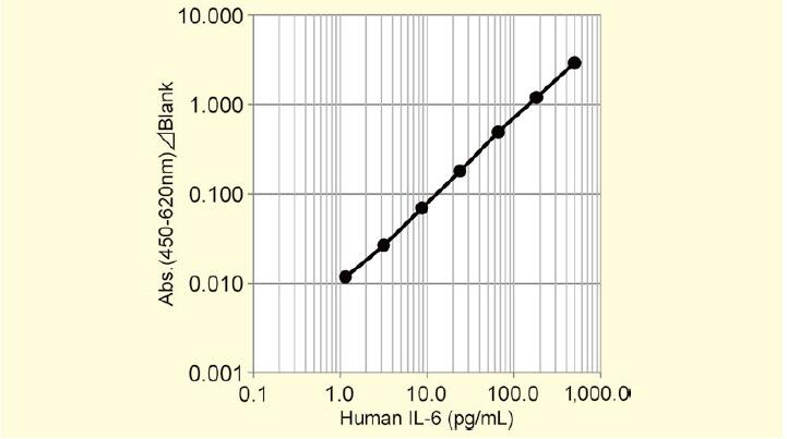
◆Lbis® Human IL-6 ELISA Kit


  IL-6是189个氨基酸的分泌性糖蛋白,是促进B细胞分化成抗体生成细胞的细胞因子。有研究表示 ,IL-6与类风湿关节炎的病情有关,其作用在类风湿关节炎等自身免疫性疾病、炎症性疾病领域受到关注。

  本试剂盒能短时间,高灵敏度检测人血清(血浆)中的微量IL-6。

产品概要

● 标准曲线范围:1.16~500pg/mL

● 检测时间:总反应时间3小时50分

● 样本量:100μL

● 测定波长:主波长450nm/副波长620nm

● 样本:人血清/血浆(肝素/EDTA)


〈标准曲线〉

增加3种产品! 可检测血清血浆样本Lbis® 试剂盒


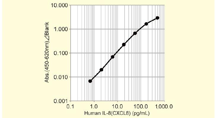
◆Lbis® Human IL-8(CXCL8)ELISA Kit


  IL-8是通过炎症性细胞因子的刺激在成纤维细胞或单核细胞、血管内皮细胞中产生的72或77个氨基酸的2种类型的炎症性CXC趋化因子。IL-8与多种疾病的相关,并在类风湿关节炎、哮喘等呼吸道疾病、痛风、牙周炎、癌症等研究领域受到了关注。

  本试剂盒能短时间,高灵敏度检测人血清(血浆)中的微量IL-8。

产品概要

● 标准曲线范围:0.686~500pg/mL

● 检测时间:总反应时间3小时50分

● 样本量:100μL

● 测定波长:主波长450nm/副波长620nm

● 样本:人血清/血浆(肝素/EDTA)


〈标准曲线〉

增加3种产品! 可检测血清血浆样本Lbis® 试剂盒


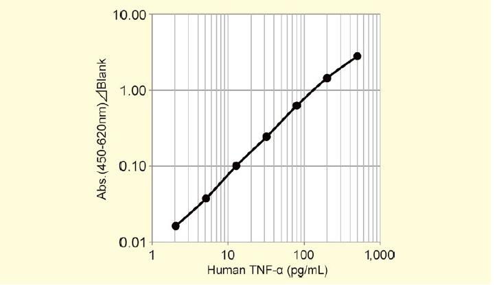
◆Lbis® Human TNF-α ELISA Kit


  TNF-α是能引起移植到小鼠中的肿瘤发生出血性坏死的诱导因子,是由157个氨基酸组成的炎症性细胞因子。TNF-α与多种疾病相关,在类风湿关节炎、炎症、糖尿病・高血脂、肾病、白血病、骨质疏松等领域受到关注。

  本试剂盒能短时间,高灵敏度检测人血清(血浆)中的微量TNF-α。


产品概要

● 标准曲线范围:2.05~500pg/mL

● 检测时间:总反应时间3小时50分

● 样本量:100μL

● 测定波长:主波长450nm/副波长620nm

● 样本:人血清/血浆(肝素/EDTA)


〈标准曲线〉

增加3种产品! 可检测血清血浆样本Lbis® 试剂盒


欲了解相关信息请点击文字:

新产品 人IL-6/IL-8/TNF-α ELISA试剂盒发售通知

Lbis® 疾病相关动物模型ELISA试剂盒系列

产品编号 产品名称 产品规格 产品等级 产品价格
635-42311 人IL-6 ELISA试剂盒,AKH-IL6
LBIS Human IL-6 ELISA Kit
96次
632-42321 人IL-8(CXCL8) ELISA试剂盒,AKH-IL8
LBIS Human IL-8(CXCL8) ELISA Kit
96次
639-42331 人 TNF-α ELISA试剂盒,AKH-TNFA
LBIS Human TNF-α ELISA Kit
96次

Nalgene 4252-0065粉体漏斗 68ml 聚丙烯PP漏斗 顶部内径65mm4252-0100 243ml-赛默飞中国代理商

产品信息
产品名称:
Nalgene 4252-0065粉体漏斗 68ml 聚丙烯PP漏斗 顶部内径65mm
产品型号:
4252-0100 243ml
Nalgene 4252-0065粉体漏斗 68ml 聚丙烯PP漏斗 顶部内径65mm4252-0100 243ml 产品特点
  Nalgene 4252-0065粉体漏斗,聚丙烯,漏斗顶部内径65mm FUNNEL POWDER PP;65MM● 聚丙烯材质● 用于传输粉末● 平行漏嘴可Z大限度地减低粉末沿黏管壁● 外部肋材可防止气塞● 传输速度快且效率高● 可高温高压灭菌

Nalgene 4252-0065粉体漏斗 68ml 聚丙烯PP漏斗 顶部内径65mm4252-0100 243ml
产品详细信息:

Nalgene 4252-0065粉体漏斗,聚丙烯,漏斗顶部内径65mm FUNNEL POWDER PP;65MM

● 聚丙烯材质
● 用于传输粉末
● 平行漏嘴可zui大限度地减低粉末沿黏管壁
● 外部肋材可防止气塞
● 传输速度快且效率高
● 可高温高压灭菌

Whatman10401631混合纤维素酯膜 ME25 0.45um 142MM 25/PK

【简单介绍】

Whatman混合纤维素酯膜是由20%的醋酸纤维素和80%的硝酸纤维素组成。与纯硝酸纤维素膜相比,这种膜的表面更加光滑和均匀,而且为颗粒检测提供了良好的颜色对比,将眼睛疲劳减至最小程度。

【简单介绍】

Whatman混合纤维素酯膜是由20%的醋酸纤维素和80%的硝酸纤维素组成。与纯硝酸纤维素膜相比,这种膜的表面更加光滑和均匀,而且为颗粒检测提供了良好的颜色对比,将眼睛疲劳减至最小程度。

【详细说明】

原装进口英国Whatman10401631混合纤维素酯膜 ME25 0.45um 142MM 25/PK

英国Whatman10401631混合纤维素酯膜 ME25 0.45um 142MM 25/PK

Membra-Fil®混酯膜

Whatman混合纤维素酯膜是由20%的醋酸纤维素和80%的硝酸纤维素组成。与纯硝酸纤维素膜相比,这种膜的表面更加光滑和均匀,而且为颗粒检测提供了良好的颜色对比,将眼睛疲劳减至最小程度。

光滑或网格

许多微生物技术包括培养后的克隆计数作为定量的标准方法。Whatman网格滤膜内有间隔3.1mm的网格线。墨水是无毒的,并且完全不含细菌生长的抑制剂

Whatman黑色混酯膜中,平滑型的用于自动克隆计数应用,而网格的则用于人工计数。黑膜使得残留物或细胞颜色和膜之间颜色有差异,不必对膜作染色处理。

无菌膜

对于那些选择使用通过消毒的灭菌了的膜的微生物工作,Whatman提供用于实验消毒的黑色网格膜,包装附有垫片。

特征和优点:

?有消毒了的膜可供主要实验选择;

?非常好的对比,方便颗粒探测;

?网格没有毒性,也不会抑制细菌生长,保证样品的完整性;

?消毒后可重复使用;

?黑色平滑或黑色网格膜中硝酸纤维素和醋酸纤维素的比例为8020

?膜具有高内部面积适合吸附力高的样品;

?更高的灰尘负载力

?低蛋白吸附特性;

?生物学惰性,好的热稳定性;

?没有表面活性剂污染样品

?膜均匀的微孔结构提高了流速

应用:

这个膜在要求更高流速和更大过滤容量的应用中非常有效,包括水溶液的澄清或消毒、分析和去除颗粒、空气中监测和微生物分析,其他应用包括:

?水溶液的澄清或消毒

?细胞学

?空气监测

?HPLC样品制备(水样)

?病毒浓缩

?微生物分析

?食物行业中,食物微生物学包括E.coil计数

?细菌生物学研究

?液态和气态颗粒计数

?酵母和霉菌

产品选择混酯膜

型号

厚度(µm

水流速

Δp=0.9mbar

(mL/min/cm2)

空气流速

Δp=3mbar

(mL/min/cm2)

起泡点

bar

WME

140

ME 24

135

25

3.7

ME 25

135

45

25

2.5

ME 26

135

110

45

1.5

ME 27

140

170

80

1.3

ME 28

140

240

100

0.8

ME 29

150

400

140

0.7

订货信息混酯膜

尺寸(mm

孔径(µm

货号

型号

无菌

数量/

ME 25

90

0.45

10401618

平滑

50

100

0.45

10401621

平滑

50

110

0.45

10401626

平滑

50

142

0.45

10401631

平滑

25

*不含交错垫片

上海金畔生物科技有限公司

文章号19742481-19742481

和光纯药的细胞因子

和光纯药的细胞因子

  • 产品特性
  • 相关资料
  • Q&A
  • 参考文献

和光纯药的细胞因子和光纯药的细胞因子

种类丰富


  提供众多种类人,小鼠,大鼠的重组细胞因子和生长因子等,具体请参阅产品列表。

  各种细胞因子的生物活性等产品详情、大包装的价格、货期请咨询。


◆产品介绍


bFGF Solution, MF

  bFGF【成纤维细胞生长因子(碱性)】是人类ES / iPS细胞的维持培养所必需的因子。也用于心肌细胞等诱导分化。本产品是无菌溶液,可直接在培养基中添加使用。同时,已注册原药等登记台账(Master File:MF)。

  bFGF广泛存在于胎儿期和成人体内的各种组织。能促进培养血管内皮细胞的增殖、迁移、蛋白酶的生产、管腔形成等。在体内具有血管生成作用。除了作为血管生成因子外,也有研究表明其在骨和软骨形成、中枢神经系统等方面具有各种生物学作用。

● 内毒素:≦10EU/mg

● 活性:≧500,000IU/mg

● 表达细胞:E. coli

● 形状:溶液型 无载体Carrier free

DKK-1, Human, recombinant

  DKK-1是Wnt/β-catenin信号通路拮抗剂。由形成低密度脂蛋白(LDL)受体相关蛋白5(LRP5)和LRP6形成复合体、抑制Wnt和Frizzled(Fz)的相互作用来抑制Wnt信号。本产品是成熟型的人DKK-1的235个氨基酸残基构成的糖蛋白。

● 内毒素≦0.1ng/μg

● 活性根据HCT116结肠癌细胞的增殖抑制功能来决定(DKK-1浓度为200ng/mL时能达到约40%的生长抑制)

● 表达细胞:HEK293 cells

● 形状:冻干品,无载体,Carrier free

LIF, Human, recombinant, Animal-derived-free

  本产品在培养和纯化的生产过程中不使用动物源性物质。

  LIF(Leukemia Inhibitory Factor)可抑制白血病细胞增殖,诱导分化巨噬细胞的因子。其他还具有参与神经元分化,骨形成,脂肪细胞的脂质运输,肾上腺皮质激素的产生等众多的功能。同时,LIF具有抑制小鼠ES细胞分化的作用,能维持ES细胞的未分化状态,被应用于细胞培养。本产品是由180个氨基酸残基构成的蛋白质。


● 内毒素≦0.01ng/μg
● 活性ED50:0.1ng/mL以下(根据人TF-1细胞的增殖刺激能力决定)

● 比活性>1×10⁷units/mg)

● 表达细胞:E. coli

● 形状:冻干品,无载体,Carrier free

Sonic Hedgehog, Human, recombinant

  Sonic Hedgehog是Hedgehog Family 5种蛋白质中的其中一种,是分泌型的信号肽。在激活分化诱导活性的位点发现Sonic Hedgehog表达,发育过程中主要参与控制分化和形态的调控。

● 内毒素≦0.1ng/μg

● 活性ED50:0.8~1.0μg/mL(根据具有碱性磷酸酶产生的C3H/10T1/2(CCL-226)细胞的诱导能力决定)

● 表达细胞:E. coli

● 形状:冻干品,无载体,Carrier free

Bone Morphogenetic Protein 7;BMP-7(骨形态发生蛋白7,人,重组)

  骨形态发生蛋白7(Bone Morphogenetic Protein 7;BMP-7)是属于TGF-β超家族的生长因子。亦称为骨诱导因子-1 (Osteogenic Protein-1;OP-1)。与特定的骨诱导载体如胶原蛋白等一起用于骨缺损治疗的应用研究。BMP-7在发育组织中广泛表达,研究发现,BMP-7基因敲除小鼠的眼、肾脏、骨架形成等出现异常,出生后容易致死。


Interleukin-6;IL-6(可溶性白细胞介素-6受体α,人,重组)

  白细胞介素 – 6(Interleukin-6;IL-6)是一种与B细胞活化等相关的炎性细胞因子。IL-6与IL-6受体α(IL-6Rα)形成复合物并与gp130结合以在细胞内发出信号。本产品在细胞外区域,低浓度下可作为IL-6活性的激动剂。


大包装

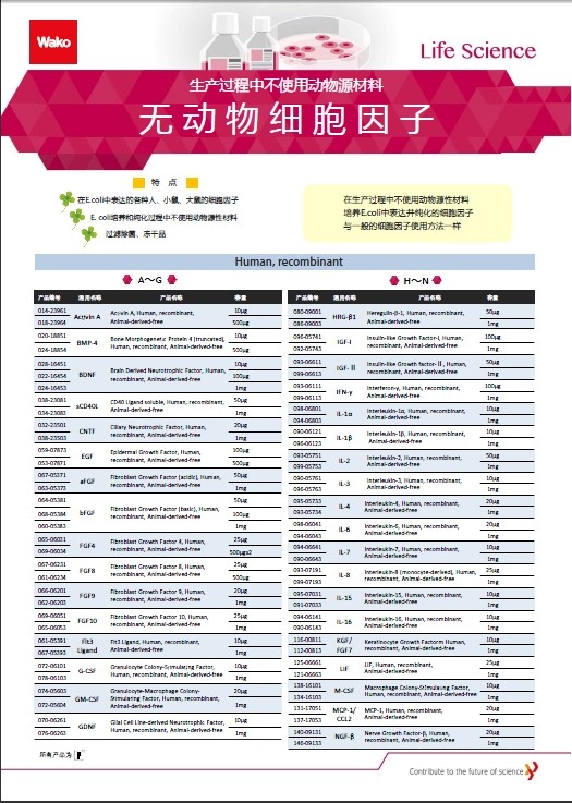
  我们提供多款细胞因子大包装规格。使用量大的客户可考虑购买。同时,还有多种无动物源细胞因子产品。

  ※无动物源细胞因子是在生产过程中不使用动物来源的原料,利用E. coli (大肠杆菌)表达纯化的细胞因子。该产品可有效降低动物引起的病毒污染的风险。

相关资料

和光纯药的细胞因子 和光纯药的细胞因子
细胞因子▪生长因子
无动物细胞因子

产品编号 产品名称 产品规格 产品等级 产品价格
062-06661 bFGF Solution, MF 50μl
068-06663 bFGF Solution, MF 50μlx4
044-34231 DKK-1, Human, recombinant 10μg
040-34233 DKK-1, Human, recombinant 1mg
125-06661 LIF, Human, recombinant, Animal-derived-free 25μg
121-06663 LIF, Human, recombinant, Animal-derived-free 1mg
198-18341 Sonic Hedgehog, Human, recombinant 25μg
194-18343 Sonic Hedgehog, Human, recombinant 500μg
014-23961 Activin A, Human, recombinant, Animal-derived-free 10μg
018-23964 Activin A, Human, recombinant, Animal-derived-free 500μg
020-18851 Bone Morphogenetic Protein 4 (truncated),
 Human, recombinant, Animal-derived-free
10μg
024-18854 Bone Morphogenetic Protein 4 (truncated),
 Human, recombinant, Animal-derived-free
500μg
028-16451 Brain Derived Neurotrophic Factor, Human, recombinant,
 Animal-derived-free
10μg
022-16454 Brain Derived Neurotrophic Factor, Human, recombinant,
 Animal-derived-free
100μg
024-16453 Brain Derived Neurotrophic Factor, Human, recombinant,
 Animal-derived-free
1mg
059-07873 Epidermal Growth Factor, Human, recombinant, 
Animal-derived-free
100μg
053-07871 Epidermal Growth Factor, Human, recombinant, 
Animal-derived-free
500μg
067-05371 Fibroblast Growth Factor (acidic), Human, recombinant, Animal-derived-free 50μg
063-05373 Fibroblast Growth Factor (acidic), Human, recombinant, Animal-derived-free 1mg
064-05381 Fibroblast Growth Factor (basic), Human, recombinant 50μg
068-05384 Fibroblast Growth Factor (basic), Human, recombinant 100μg
060-05383 Fibroblast Growth Factor (basic), Human, recombinant 1mg
064-04541 Fibroblast Growth Factor (basic), Human, recombinant, Animal-derived-free 50μg
060-04543 Fibroblast Growth Factor (basic), Human, recombinant, Animal-derived-free 100μg
068-04544 Fibroblast Growth Factor (basic), Human, recombinant, Animal-derived-free 1mg
067-06231 Fibroblast Growth Factor 8, Human, recombinant, Animal-derived-free 25μg
061-06234 Fibroblast Growth Factor 8, Human, recombinant, Animal-derived-free 500μg
061-05391 Flt3 Ligand, Human, recombinant, Animal-derived-free 10μg
067-05393 Flt3 Ligand, Human, recombinant, Animal-derived-free 1mg
070-06261 Glial Cell Line-derived Neurotrophic Factor,
Human, recombinant,  Animal-derived-free
10μg
076-06263 Glial Cell Line-derived Neurotrophic Factor,
Human, recombinant,  Animal-derived-free
1mg
119-00661 Keratinocyte Growth Factorm Human, recombinant 10μg
116-00811 Keratinocyte Growth Factorm Human, recombinant,  Animal-derived-free 10μg
112-00813 Keratinocyte Growth Factorm Human, recombinant,  Animal-derived-free 1μg
146-09231 Neurotrophin-3, Human, recombinant, Animal-derived-free 10μg
142-09233 Neurotrophin-3, Human, recombinant, Animal-derived-free 1mg
197-15511 Stem Cell Factor, Human, recombinant, Animal-derived-free 10μg
191-15514 Stem Cell Factor, Human, recombinant, Animal-derived-free 250μg
193-15513 Stem Cell Factor, Human, recombinant, Animal-derived-free 1mg
207-19281 Transforming Growth Factor-β1, Human, recombinant 10μg
203-19283 Transforming Growth Factor-β1, Human, recombinant 1mg
207-19281 Transforming Growth Factor-β3, 
Human, recombinant,  Animal-derived-free
10μg
203-19283 Transforming Growth Factor-β3, 
Human, recombinant,  Animal-derived-free
1mg
203-15263 Tumor Necrosis Factor-α, Human, recombinant 10μg
207-15261 Tumor Necrosis Factor-α, Human, recombinant 50μg
201-15264 Tumor Necrosis Factor-α, Human, recombinant 1mg
207-17581 Thrombopoietin, Human, recombinant, Animal-derived-free 10μg
201-17584 Thrombopoietin, Human, recombinant, Animal-derived-free 500μg
203-17583 Thrombopoietin, Human, recombinant, Animal-derived-free 1mg
223-01311 Vascular Endothelial Growth Factor-A165, Human, recombinant 10μg
229-01313 Vascular Endothelial Growth Factor-A165, Human, recombinant 1mg
226-01781 Vascular Endothelial Growth Factor-A165,
 Human, recombinant,  Animal-derived-free
10μg
226-01786 Vascular Endothelial Growth Factor-A165,
 Human, recombinant,  Animal-derived-free
100μg
220-01784 Vascular Endothelial Growth Factor-A165,
 Human, recombinant,  Animal-derived-free
500μg
222-01783 Vascular Endothelial Growth Factor-A165,
 Human, recombinant,  Animal-derived-free
1mg
026-19171 Bone Morphogenetic Protein 7(BMP-7 / BMP7 / OP-1), Human, recombinant (expressed in CHO Cells) 骨形态生成蛋白7(BMP-7、BMP7 / OP-1),人,重组 (在CHO细胞中表达) 10 ug
092-07301 Interleukin-6 Receptor α soluble(sIL-6Ralpha), Human, recombinant 白细胞介素-6受体α可溶性(sIL-6ralpha),人,重组 20μg

普通MA板无菌箱ST-2型日本三博特sanplatec

普通MA板无菌箱ST-2型
产品编号: WEB0187 价格: 会员价:0元;市场价:0元 产品特点
产品规格

标准培植< ?xml:namespace prefix = o ns = "urn:schemas-microsoft-com:office:office" />

手套1

规格(mm)

800(W)×550(D)×500(H)

材料

确认材料的耐药性 >> 耐药性检索

        普通MA板无菌箱ST-2型普通MA板无菌箱ST-2型产品特征

开口部大,使用方便。

箱门有锁,安全放心。

可选择客人需要的部件,更简便经济。(从无菌箱用部件里选择)

可订做特殊规格,详情请咨询。


图纸

请点击查看。


StemSure® hPSC去除剂【rBC2LCN-PE38】

StemSure® hPSC去除剂【rBC2LCN-PE38】

  • 产品特性
  • 相关资料
  • Q&A
  • 参考文献

StemSure® hPSC去除剂【rBC2LCN-PE38】人ES/iPS细胞强力去除剂

StemSure® hPSC去除剂【rBC2LCN-PE38】


  rBC2LCN(AiLecS1) 是Burkholderiacenocepacia源凝聚素BC2L-C的N末端域在大肠杆菌表达的重组凝聚素。rBC2LCN对存在于人ES/iPS细胞表面的糖链具有非常高的特异性。

  本品是使绿脓杆菌源外毒素的位置域(38kDa)融合在rBC2LCN的N末端部分的重组蛋白。通过进入细胞内引起细胞死亡,选择性地去除人ES/iPS细胞。其活性比具有相同作用的rBC2LCN-PE23高强百倍。


◆特点


● 在分化诱导时能选择性地去除残留的人ES/iPS细胞

● 将本品添加至培养液中,即可简单高效地去除培养液中的人ES/iPS细胞

● 原料中不含动物源成分


数据


去除人iPS细胞

StemSure® hPSC去除剂【rBC2LCN-PE38】


1. StemSure® hPSC去除剂

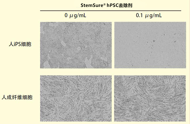
  在人iPS细胞201B7细胞株和人成纤维细胞的培养液中添加StemSure® hPSC去除剂(终浓度0.1μg/ mL),培养48h,换液后再培养24h。结果显示,用StemSure® hPSC去除剂处理过的人iPS细胞基本上都已被去除(右上)。用同样的方法处理人成纤维细胞,则完全没有被去除(右下)。

产品列表


产品编号

产品名称

规格

容量

199-18511

195-18513

StemSure® hPSC去除液

【rBC2LCN-PE38】

StemSure® hPSC Remover

【rBC2LCN-PE38】

细胞培养用

100μL

100μL×5


◆相关产品


产品编号

产品名称

规格

容量

029-18061

025-18063

BC2LCN【AiLecS1】凝集素,重组,溶液

BC2LCN【AiLecS1】 Lectin, recombinant, Solution

糖链研究用

1mg

1mg×5

180-02991

186-02993

人iPS未分化标记染料rBC2LCN-FITC 

【AiLecS1-FITC】

rBC2LCN-FITC【AiLecS1-FITC】

Excitation 495nm, Emission 520nm

细胞染色用

100μL

100μL×5

186-03211

182-03213

人iPS未分化标记染料rBC2LCN-547

【AiLecS1-547】

rBC2LCN-547【AiLecS1-547】

Excitation 551nm, Emission 565nm

细胞染色用

100μL

100μL×5

185-03161

181-03163

人iPS未分化标记染料rBC2LCN-635 

【AiLecS1-635】

rBC2LCN-635【AiLecS1-635】

Excitation 634nm, Emission 654nm

细胞染色用

100μL

100μL×5

180-03231

186-03233

人ES/iPS细胞清除试剂

rBC2LCN-PE23

细胞培养用

100μL

100μL×5

299-78301

人类ES / iPS细胞监测试剂盒

Human ES/iPS Cell Monitoring Kit

再生医疗研究用

96 次


[1] Tateno, H., Onuma, Y., Ito, Y., Minoshima, F., Saito, S., Shimizu, M., Aiki, Y., 

   Asashima, M. and Hirabayashi, J. : Stem Cell Reports , 4 , 811( 2015).

[2] Tateno, H., Minoshima, F. and Saito, S. : Molecules , 22 , 1151( 2017)

产品编号 产品名称 产品规格 产品等级 产品价格

布氏漏斗,PP材质, 布氏漏斗 PP漏斗 塑料漏斗 供应

  • 产品描述

布氏漏斗是实验室中使用的一种塑料仪器用来使用真空或负压力抽吸进行过滤。
用于真空过滤,提取结晶等
多种规格可选:60-150mm

· 两件式,易于清洗

· 适配标准滤纸

产品编号 

顶部内径

(mm)

包装形式

84110-1001

84110-1002

84110-1003

84110-1004

84110-1005

60

75

95

130

150

1只/袋, 8只/箱

1只/袋, 6只/箱

1只/袋, 4只/箱

1只/袋, 4只/箱

1只/袋, 4只/箱

Phos-tag™ 质谱分析试剂盒

Phos-tag™ 质谱分析试剂盒
Phos-tag™ Mass Analytical Kit

  • 产品特性
  • 相关资料
  • Q&A
  • 参考文献

Phos-tag™ Mass Analytical KitPhos-tag™ 质谱分析试剂盒

用于MALDI-TOF/MS检测,提高检测灵敏度!

  用于质谱分析的试剂套装。

  Phos-tag Mass Analytical Kit 是用于质谱分析的试剂套装,可配套MALDI-TOF/MS使用。可检测磷酸化分子- Phos-tag® 复合物,通常可提高低磷酸化分子的检测灵敏度。


试剂盒内容:

● Phos-tag™ MS-101L  5 mg([C27H29N6O64Zn2]3+ MW:581.4)

● Phos-tag™ MS-101H 5 mg([C27H29N6O68Zn2]3+ MW:589.4)

● Phos-tag™ MS-101N 10 mg([C27H29N6OZn2]3+ MW:584.3)

原理:


Phos-tag™ 质谱分析试剂盒


优点、特色:

● CH3COO- 等价结合在Phos-tag™ MS-101上。

● 在溶液中,不含有阴离子的Phos-tag™ MS-101 带有+3价。

● 检测前需制备1mM 的Phos-tag™ MS-101L,MS-101H或者MS-101N( 溶于水)。

案例、应用:

【使用例子:检测Phos-tag™ – 磷酸化LPA 复合体】


Phos-tag™ 质谱分析试剂盒


由于正电荷增大磷酸化LPA 检测灵敏度上升


Phos-tag™ 系列

磷酸化蛋白新方法!

  Phos-tag™ 是一种能与磷酸离子特异性结合的功能性分子。它可用于磷酸化蛋白的分离(Phos-tag™ Acrylamide)、Western Blot检测(Phos-tag™ Biotin)、蛋白纯化 (Phos-tag™ Agarose)及质谱分析MALDI-TOF/MS (Phos-tag™ Mass Analytical Kit)。


Phos-tag™ 的基本结构:

Phos-tag™ 质谱分析试剂盒

特点:

与-2价磷酸根离子的亲和性和选择性高于其它阴离子

在pH 5-8的生理环境下生成稳定的复合物

原理:


Phos-tag™ 质谱分析试剂盒

相关应用:


Phos-tag™ 质谱分析试剂盒

相关产品:

 产品名称

 用  途

 Phos-tag™ Acrylamide

 分离: SDS – PAGE 分离不同磷酸化水平的蛋白

 SuperSep Phos-tag™

 分离: 预制胶中含有50μM Phos-tag™ Acrylamide

 Phos-tag™ Biotin

 检测: 代替 Western Blot 检测中的磷酸化抗体

 Phos-tag™ Agarose

 纯化: 通用柱层析,纯化磷酸化蛋白

 Phos-tag™ Mass

 Analytical Kit

 分析: 用于质谱 MALDI-TOF/MS 分析,提高磷酸化分子的检测灵敏度


phos-tag™由日本广岛大学研究生院医齿药学综合研究科医药分子功能科学研究室开发。

更多产品信息,请点击:http://phos-tag.jp

Phos-tag™ 质谱分析试剂盒

Phos-tag 第5版说明书

Phos-tag™ 质谱分析试剂盒

Phos-tag系列 ver 5

Q.     Phos-tag™ Mass 用于实验可以使用多少次?

A.     如果每次用量为5μL,至少可以使用1000 次。

Q.     如何选择使用Phos-tag™ MS-101L,Phos-tag™ MS-101H 和Phos-tag™ MS-101N ?

A.     Phos-tag™ 101N 含有自然存在的Zn,101L 与101H 分别含有Zn 的同位素64Zn 和68Zn。

     请参考以下建议:

     摸索条件时使用101N,其中含有多种同位素,结果比较详细;

     鉴定磷酸基团是否存在,使用101L 和101H,这些试剂分别包含64Zn 和68Zn。使用这些试剂检测同一个样品

     时会产生不同的荷质比。

Q.     如果想测定经过Phos-tag™ SDS-PAGE 分离得到的样品,是否必须要在凝胶消化之前去除Phos-tag™?

A.     没有必要。SDS-PAGE 结束之后根据一般的凝胶消化方法进行操作即可。

Q.     能否用于ESI 质谱?

A.     是的,可以使用。请参考下面的文献,这篇报道使用Phos-tag™ MS-101N 进行ESI-MS 分析。在实验过程

     中,使用了中性溶液,若为酸性溶液会导致Phos-tag ™ 分离。

        【参考文献】 Anal. Chem. (2008), 80, 2531-2538 (MS-101N ESI-MS)

【参考文献】


·  Conversion of graded phosphorylation into switch-like nuclear translocation via autoregulatory mechanisms in ERK signalling[J].Nature communications, 2016, 7,Shindo Y, Iwamoto K, Mouri K, et al.

·  PTEN modulates EGFR late endocytic trafficking and degradation by dephosphorylating Rab7[J]. Nature communications, 2016, 7,Shinde S R, Maddika S.

·  Feedback control of ErbB2 via ERK-mediated phosphorylation of a conserved threonine in the juxtamembrane domain[J]. Scientific Reports, 2016, 6: 31502,Kawasaki Y, Sakimura A, Park C M, et al.

·  Plastid-nucleus communication involves calcium-modulated MAPK signalling[J]. Nature Communications, 2016, 7,Guo H, Feng P, Chi W, et al.

·  Sequential domain assembly of ribosomal protein S3 drives 40S subunit maturation[J]. Nature communications, 2016, 7,Mitterer V, Murat G, Réty S, et al.

·  Phos-tag analysis of Rab10 phosphorylation by LRRK2: a powerful assay for assessing kinase function and inhibitors[J]. Biochemical Journal, 2016: BCJ20160557,Ito G, Katsemonova K, Tonelli F, et al.

·  Analysis of phosphorylation of the myosin targeting subunit of smooth muscle myosin light chain phosphatase by Phos-tag SDS-PAGE[J]. The FASEB Journal, 2016, 30(1 Supplement): 1209.1-1209.1,Walsh M P, MacDonald J A, Sutherland C.

·  Using Phos-Tag in Western Blotting Analysis to Evaluate Protein Phosphorylation[J]. Kidney Research: Experimental Protocols, 2016: 267-277,Horinouchi T, Terada K, Higashi T, et al.

·  The Abundance of Nonphosphorylated Tau in Mouse and Human Tauopathy Brains Revealed by the Use of Phos-Tag Method[J]. The American journal of pathology, 2016, 186(2): 398-409,Kimura T, Hatsuta H, Masuda-Suzukake M, et al.

·  Phos-tag SDS-PAGE resolves agonist-and isoform-specific activation patterns for PKD2 and PKD3 in cardiomyocytes and cardiac fibroblasts[J]. Journal of Molecular and Cellular Cardiology, 2016,Qiu W, Steinberg S F.

·  Analysis of phosphorylation of the myosin-targeting subunit of myosin light chain phosphatase by Phos-tag SDS-PAGE[J]. American Journal of Physiology-Cell Physiology, 2016, 310(8): C681-C691,Sutherland C, MacDonald J A, Walsh M P.

·  Electrochemical biosensor for protein kinase A activity assay based on gold nanoparticles-carbon nanospheres, phos-tag-biotin and β-galactosidase[J]. Biosensors and Bioelectronics, 2016, 86: 508-515,Zhou Y, Yin H, Li X, et al.

·  Validation of Cis and Trans Modes in Multistep Phosphotransfer Signaling of Bacterial Tripartite Sensor Kinases by Using Phos-Tag SDS-PAGE[J]. PloS one, 2016, 11(2): e0148294,Kinoshita-Kikuta E, Kinoshita E, Eguchi Y, et al.

·  Phosphopeptide Detection with Biotin-Labeled Phos-tag[J]. Phospho-Proteomics: Methods and Protocols, 2016: 17-29,Kinoshita-Kikuta E, Kinoshita E, Koike T.

·  A Phos‐tag SDS‐PAGE method that effectively uses phosphoproteomic data for profiling the phosphorylation dynamics of MEK1[J]. Proteomics, 2016,Kinoshita E, Kinoshita‐Kikuta E, Kubota Y, et al.

·  Difference gel electrophoresis of phosphoproteome: U.S. Patent Application 15/004,339[P]. 2016-1-22,Tao W A, Wang L.

·  ERK1/2-induced phosphorylation of R-Ras GTPases stimulates their oncogenic potential[J]. Oncogene, 2016,Frémin C, Guégan J P, Plutoni C, et al.

·  Microtubules Inhibit E-Cadherin Adhesive Activity by Maintaining Phosphorylated p120-Catenin in a Colon Carcinoma Cell Model[J]. PloS one, 2016, 11(2): e0148574,Maiden S L, Petrova Y I, Gumbiner B M.

·  Serine 231 and 257 of Agamous-like 15 are phosphorylated in floral receptacles[J]. Plant Signaling & Behavior, 2016, 11(7): e1199314,Patharkar O R, Macken T A, Walker J C.

·  A small molecule pyrazolo [3, 4-d] pyrimidinone inhibitor of zipper-interacting protein kinase suppresses calcium sensitization of vascular smooth muscle[J]. Molecular pharmacology, 2016, 89(1): 105-117,MacDonald J A, Sutherland C, Carlson D A, et al.

·  The RNA polymerase II C-terminal domain phosphatase-like protein FIERY2/CPL1 interacts with eIF4AIII and is essential for nonsense-mediated mRNA decay in Arabidopsis[J]. The Plant Cell, 2016: TPC2015-00771-RA,Chen T, Qin T, Ding F, et al.

·  Vasorelaxant Effect of 5′-Methylthioadenosine Obtained from Candida utilis Yeast Extract through the Suppression of Intracellular Ca2+ Concentration in Isolated Rat Aorta[J]. Journal of agricultural and food chemistry, 2016, 64(17): 3362-3370,Kumrungsee T, Akiyama S, Saiki T, et al.

·  Inhibition of deubiquitinating activity of USP14 decreases tyrosine hydroxylase phosphorylated at Ser19 in PC12D cells[J]. Biochemical and biophysical research communications, 2016, 472(4): 598-602,Nakashima A, Ohnuma S, Kodani Y, et al.

·  Actin Tyrosine-53-Phosphorylation in Neuronal Maturation and Synaptic Plasticity[J]. The Journal of Neuroscience, 2016, 36(19): 5299-5313,Bertling E, Englund J, Minkeviciene R, et al.

·  AMPK-dependent phosphorylation of lipid droplet protein PLIN2 triggers its degradation by CMA[J]. Autophagy, 2016, 12(2): 432-438,Kaushik S, Cuervo A M.

·  Myocardin-related transcription factor a and yes-associated protein exert dual control in G protein-coupled receptor-and RhoA-mediated transcriptional regulation and cell proliferation[J]. Molecular and cellular biology, 2016, 36(1): 39-49,Olivia M Y, Miyamoto S, Brown J H.

·  Extensive phosphorylation of AMPA receptors in neurons[J]. Proceedings of the National Academy of Sciences, 2016, 113(33): E4920-E4927,Diering G H, Heo S, Hussain N K, et al.

·  The transmembrane region of guard cell SLAC1 channels perceives CO2 signals via an ABA-independent pathway in Arabidopsis[J]. The Plant Cell, 2016, 28(2): 557-567,Yamamoto Y, Negi J, Wang C, et al.

·  The Hippo pathway mediates inhibition of vascular smooth muscle cell proliferation by cAMP[J]. Journal of molecular and cellular cardiology, 2016, 90: 1-10,Kimura T E, Duggirala A, Smith M C, et al.

·  Atg13 is essential for autophagy and cardiac development in mice[J]. Molecular and cellular biology, 2016, 36(4): 585-595,Kaizuka T, Mizushima N.

·  The ChrSA and HrrSA two-component systems are required for transcriptional regulation of the hemA promoter in Corynebacterium diphtheriae[J]. Journal of Bacteriology, 2016: JB. 00339-16,Burgos J M, Schmitt M P.

·  Intergenic Variable-Number Tandem-Repeat Polymorphism Upstream of rocA Alters Toxin Production and Enhances Virulence in Streptococcus pyogenes[J]. Infection and Immunity, 2016, 84(7): 2086-2093,Zhu L, Olsen R J, Horstmann N, et al.

·  Receptor for advanced glycation end products (RAGE) knockout reduces fetal dysmorphogenesis in murine diabetic pregnancy[J]. Reproductive Toxicology, 2016, 62: 62-70,Ejdesjö A, Brings S, Fleming T, et al.

·  Aurora kinase-induced phosphorylation excludes transcription factor RUNX from the chromatin to facilitate proper mitotic progression[J]. Proceedings of the National Academy of Sciences, 2016, 113(23): 6490-6495,Chuang L S H, Khor J M, Lai S K, et al.

·  Quantitative phosphoproteomics of protein kinase SnRK1 regulated protein phosphorylation in Arabidopsis under submergence[J]. Journal of experimental botany, 2016: erw107,Cho H Y, Wen T N, Wang Y T, et al.

·  Temporal regulation of lipin activity diverged to account for differences in mitotic programs[J]. Current Biology, 2016, 26(2): 237-243,Makarova M, Gu Y, Chen J S, et al.

·  Block of CDK1‐dependent polyadenosine elongation of Cyclin B mRNA in metaphase‐i‐arrested starfish oocytes is released by intracellular pH elevation upon spawning[J]. Molecular reproduction and development, 2016, 83(1): 79-87,Ochi H, Aoto S, Tachibana K, et al.

·  Mitotic Exit Function of Polo-like Kinase Cdc5 Is Dependent on Sequential Activation by Cdk1[J]. Cell reports, 2016, 15(9): 2050-2062,Rodriguez-Rodriguez J A, Moyano Y, Játiva S, et al.

·  PLK2 phosphorylates and inhibits enriched TAp73 in human osteosarcoma cells[J]. Cancer medicine, 2016, 5(1): 74-87,Hu Z B, Liao X H, Xu Z Y, et al.

·  Phosphorylated TDP-43 becomes resistant to cleavage by calpain: A regulatory role for phosphorylation in TDP-43 pathology of ALS/FTLD[J]. Neuroscience research, 2016, 107: 63-69,Yamashita T, Teramoto S, Kwak S.

·  The Pch2 AAA+ ATPase promotes phosphorylation of the Hop1 meiotic checkpoint adaptor in response to synaptonemal complex defects[J]. Nucleic acids research, 2016: gkw506,Herruzo E, Ontoso D, González-Arranz S, et al.

·  An optimized guanidination method for large‐scale proteomic studies[J]. Proteomics, 2016,Ye J, Zhang Y, Huang L, et al.

·  Expression and purification of the kinase domain of PINK1 in Pichia pastoris[J]. Protein Expression and Purification, 2016,Wu D, Qu L, Fu Y, et al.

·  BRI2 and BRI3 are functionally distinct phosphoproteins[J]. Cellular signalling, 2016, 28(1): 130-144,Martins F, Rebelo S, Santos M, et al.

·  Identification of glycoproteins associated with HIV latently infected cells using quantitative glycoproteomics[J]. Proteomics, 2016,Yang W, Jackson B, Zhang H.

·  Regulation of Beclin 1 Protein Phosphorylation and Autophagy by Protein Phosphatase 2A (PP2A) and Death-associated Protein Kinase 3 (DAPK3)[J]. Journal of Biological Chemistry, 2016, 291(20): 10858-10866,Fujiwara N, Usui T, Ohama T, et al.

·  Regulatory Implications of Structural Changes in Tyr201 of the Oxygen Sensor Protein FixL[J]. Biochemistry, 2016, 55(29): 4027-4035,Yamawaki T, Ishikawa H, Mizuno M, et al.

·  Histone demethylase Jmjd3 regulates osteoblast apoptosis through targeting anti-apoptotic protein Bcl-2 and pro-apoptotic protein Bim[J]. Biochimica et Biophysica Acta (BBA)-Molecular Cell Research, 2016, 1863(4): 650-659,Yang D, Okamura H, Teramachi J, et al.

·  Analysis of Molecular Species Profiles of Ceramide-1-phosphate and Sphingomyelin Using MALDI-TOF Mass Spectrometry[J]. Lipids, 2016, 51(2): 263-270,Yamashita R, Tabata Y, Iga E, et al.

·  Highly sensitive myosin phosphorylation analysis in the renal afferent arteriole[J]. Journal of Smooth Muscle Research, 2016, 52(0): 45-55,Takeya K.

·  Functional dissection of the CroRS two-component system required for resistance to cell wall stressors in Enterococcus faecalis[J]. Journal of bacteriology, 2016, 198(8): 1326-1336,Kellogg S L, Kristich C J.

·  Regulation of mitogen-activated protein kinase by protein kinase C and mitogen-activated protein kinase phosphatase-1 in vascular smooth muscle[J]. American Journal of Physiology-Cell Physiology, 2016, 310(11): C921-C930,Trappanese D M, Sivilich S, Ets H K, et al.

·  ModProt: a database for integrating laboratory and literature data about protein post-translational modifications[J]. Journal of Electrophoresis, 2016, 60(1): 1-4,Kimura Y, Toda T, Hirano H.

·  The C-ETS2-TFEB Axis Promotes Neuron Survival under Oxidative Stress by Regulating Lysosome Activity[J]. Oxidative medicine and cellular longevity, 2016,Ma S, Fang Z, Luo W, et al.

·  Essential role of the PSI–LHCII supercomplex in photosystem acclimation to light and/or heat conditions by state transitions[J]. Photosynthesis Research, 2016: 1-10,Marutani Y, Yamauchi Y, Higashiyama M, et al.

·  Identification of a redox-modulatory interaction between selenoprotein W and 14-3-3 protein[J]. Biochimica et Biophysica Acta (BBA)-Molecular Cell Research, 2016, 1863(1): 10-18,Jeon Y H, Ko K Y, Lee J H, et al.

·  Effects of hydrogen sulfide on the heme coordination structure and catalytic activity of the globin-coupled oxygen sensor AfGcHK[J]. BioMetals, 2016, 29(4): 715-729,Fojtikova V, Bartosova M, Man P, et al.

·  Identification and functional analysis of phosphorylation in Newcastle disease virus phosphoprotein[J]. Archives of virology, 2016: 1-14,Qiu X, Zhan Y, Meng C, et al.

·  Increased level of phosphorylated desmin and its degradation products in heart failure[J]. Biochemistry and Biophysics Reports, 2016, 6: 54-62,Bouvet M, Dubois-Deruy E, Alayi T D, et al.

·  Profiling DNA damage-induced phosphorylation in budding yeast reveals diverse signaling networks[J]. Proceedings of the National Academy of Sciences, 2016: 201602827,Zhou C, Elia A E H, Naylor M L, et al.

·  Unexpected properties of sRNA promoters allow feedback control via regulation of a two-component system[J]. Nucleic Acids Research, 2016: gkw642,Brosse A, Korobeinikova A, Gottesman S, et al.

·  Evolution of ZnII–Macrocyclic Polyamines to Biological Probes and Supramolecular Assembly[J]. Macrocyclic and Supramolecular Chemistry: How Izatt-Christensen Award Winners Shaped the Field, 2016: 415,Kimura E, Koike T, Aoki S.

·  Phosphopeptide Enrichment Using Various Magnetic Nanocomposites: An Overview[J]. Phospho-Proteomics: Methods and Protocols, 2016: 193-209,Batalha Í L, Roque A C A.

·  In vivo phosphorylation of a peptide tag for protein purification[J]. Biotechnology letters, 2016, 38(5): 767-772,Goux M, Fateh A, Defontaine A, et al.

·  Regulation of cell reversal frequency in Myxococcus xanthus requires the balanced activity of CheY‐like domains in FrzE and FrzZ[J]. Molecular microbiology, 2016,Kaimer C, Zusman D R.

·  Elevation of cortical serotonin transporter activity upon peripheral immune challenge is regulated independently of p38 mitogen‐activated protein kinase activation and transporter phosphorylation[J]. Journal of neurochemistry, 2016, 137(3): 423-435,Schwamborn R, Brown E, Haase J.

·  The Yeast Cyclin-Dependent Kinase Routes Carbon Fluxes to Fuel Cell Cycle Progression[J]. Molecular cell, 2016, 62(4): 532-545,Ewald J C, Kuehne A, Zamboni N, et al.

·  Two Degradation Pathways of the p35 Cdk5 (Cyclin-dependent Kinase) Activation Subunit, Dependent and Independent of Ubiquitination[J]. Journal of Biological Chemistry, 2016, 291(9): 4649-4657,Takasugi T, Minegishi S, Asada A, et al.

·  Increased level of phosphorylated desmin and its degradation products in heart failure[J]. Biochemistry and Biophysics Reports. 2016,Bouvet M, Dubois-Deruy E, Alayi T D, et al.

·  a high‐affinity LCO‐binding protein of Medicago truncatula, interacts with LYK3, a key symbiotic receptor[J]. FEBS letters, 2016, 590(10): 1477-1487,Fliegmann J, Jauneau A, Pichereaux C, et al. LYR3,

·  Nek1 Regulates Rad54 to Orchestrate Homologous Recombination and Replication Fork Stability[J]. Molecular Cell, 2016,Spies J, Waizenegger A, Barton O, et al.

·  PhostagTM-gel retardation and in situ thylakoid kinase assay for determination of chloroplast protein phosphorylation targets[J]. Endocytobiosis and Cell Research, 2016, 27(2): 62-70,Dytyuk Y, Flügge F, Czarnecki O, et al.

·  Luteinizing Hormone Causes Phosphorylation and Activation of the cGMP Phosphodiesterase PDE5 in Rat Ovarian Follicles, Contributing, Together with PDE1 Activity, to the Resumption of Meiosis[J]. Biology of reproduction, 2016: biolreprod. 115.135897,Egbert J R, Uliasz T F, Shuhaibar L C, et al.

·  Newby, AC, & Bond, M.(2016). The Hippo pathway mediates inhibition of vascular smooth muscle cell proliferation by cAMP[J]. Journal of Molecular and Cellular Cardiology, 2016, 90: 1-10,Kimura-Wozniak T, Duggirala A, Smith M C, et al. G.

·  Yeast lacking the amphiphysin family protein Rvs167 is sensitive to disruptions in sphingolipid levels[J]. The FEBS Journal, 2016, 283(15): 2911-2928,Toume M, Tani M.

·  Regulation of CsrB/C sRNA decay by EIIAGlc of the phosphoenolpyruvate: carbohydrate phosphotransferase system[J]. Molecular microbiology, 2016, 99(4): 627-639,Leng Y, Vakulskas C A, Zere T R, et al.

·  The Late S-Phase Transcription Factor Hcm1 Is Regulated through Phosphorylation by the Cell Wall Integrity Checkpoint[J]. Molecular and cellular biology, 2016: MCB. 00952-15,Negishi T, Veis J, Hollenstein D, et al.

·  Validation of chemical compound library screening for transcriptional co‐activator with PDZ‐binding motif inhibitors using GFP‐fused transcriptional co‐activator with PDZ‐binding motif[J]. Cancer science, 2016, 107(6): 791-802,Nagashima S, Maruyama J, Kawano S, et al.

·  ULK1/2 Constitute a Bifurcate Node Controlling Glucose Metabolic Fluxes in Addition to Autophagy[J]. Molecular cell, 2016, 62(3): 359-370,Li T Y, Sun Y, Liang Y, et al.

·  Spatiotemporal dynamics of Oct4 protein localization during preimplantation development in mice[J]. Reproduction, 2016: REP-16-0277,Fukuda A, Mitani A, Miyashita T, et al.

·  The tandemly repeated NTPase (NTPDase) from Neospora caninum is a canonical dense granule protein whose RNA expression, protein secretion and phosphorylation coincides with the tachyzoite egress[J]. Parasites & Vectors, 2016, 9(1): 1,Pastor-Fernández I, Regidor-Cerrillo J, Álvarez-García G, et al.

·  Interaction Analysis of a Two-Component System Using Nanodiscs[J]. PloS one, 2016, 11(2): e0149187,Hörnschemeyer P, Liss V, Heermann R, et al.

·  Constitutive Activation of PINK1 Protein Leads to Proteasome-mediated and Non-apoptotic Cell Death Independently of Mitochondrial Autophagy[J]. Journal of Biological Chemistry, 2016, 291(31): 16162-16174,Akabane S, Matsuzaki K, Yamashita S, et al.

·  p38β Mitogen-Activated Protein Kinase Modulates Its Own Basal Activity by Autophosphorylation of the Activating Residue Thr180 and the Inhibitory Residues Thr241 and Ser261[J]. Molecular and cellular biology, 2016, 36(10): 1540-1554,Beenstock J, Melamed D, Mooshayef N, et al.

·  Lysophosphatidylcholine acyltransferase 1 protects against cytotoxicity induced by polyunsaturated fatty acids[J]. The FASEB Journal, 2016, 30(5): 2027-2039,Akagi S, Kono N, Ariyama H, et al.

·  Characterization of a herpes simplex virus 1 (HSV-1) chimera in which the Us3 protein kinase gene is replaced with the HSV-2 Us3 gene[J]. Journal of virology, 2016, 90(1): 457-473,Shindo K, Kato A, Koyanagi N, et al.

·  Generation of phospho‐ubiquitin variants by orthogonal translation reveals codon skipping[J]. FEBS letters, 2016, 590(10): 1530-1542,George S, Aguirre J D, Spratt D E, et al.

·  Evolution of KaiC-Dependent Timekeepers: A Proto-circadian Timing Mechanism Confers Adaptive Fitness in the Purple Bacterium Rhodopseudomonas palustris[J]. PLoS Genet, 2016, 12(3): e1005922,Ma P, Mori T, Zhao C, et al.

·  Phosphorylation of Bni4 by MAP kinases contributes to septum assembly during yeast cytokinesis[J]. FEMS Yeast Research, 2016, 16(6): fow060,Pérez J, Arcones I, Gómez A, et al.

·  Alteration of Antiviral Signalling by Single Nucleotide Polymorphisms (SNPs) of Mitochondrial Antiviral Signalling Protein (MAVS)[J]. PloS one, 2016, 11(3): e0151173,Xing F, Matsumiya T, Hayakari R, et al.

·  Arm-in-arm response regulator dimers promote intermolecular signal transduction[J]. Journal of bacteriology, 2016, 198(8): 1218-1229,Baker A W, Satyshur K A, Morales N M, et al.

·  The lsh/ddm1 homolog mus-30 is required for genome stability, but not for dna methylation in neurospora crassa[J]. PLoS Genet, 2016, 12(1): e1005790,Basenko E Y, Kamei M, Ji L, et al.

·  Fine tuning chloroplast movements through physical interactions between phototropins[J]. Journal of Experimental Botany, 2016: erw265,Sztatelman O, Łabuz J, Hermanowicz P, et al.

·  Characterization of the Neospora caninum NcROP40 and NcROP2Fam-1 rhoptry proteins during the tachyzoite lytic cycle[J]. Parasitology, 2016, 143(01): 97-113,Pastor-Fernandez I, Regidor-Cerrillo J, Jimenez-Ruiz E, et al.

·  Transcriptional Profile during Deoxycholate-Induced Sporulation in a Clostridium perfringens Isolate Causing Foodborne Illness[J]. Applied and environmental microbiology, 2016, 82(10): 2929-2942,Yasugi M, Okuzaki D, Kuwana R, et al.

·  Timely Closure of the Prospore Membrane Requires SPS1 and SPO77 in Saccharomyces cerevisiae[J]. Genetics, 2016: genetics. 115.183939,Paulissen S M, Slubowski C J, Roesner J M, et al.

·  DDK dependent regulation of TOP2A at centromeres revealed by a chemical genetics approach[J]. Nucleic Acids Research, 2016: gkw626,Wu K Z L, Wang G N, Fitzgerald J, et al.

·  OVATE Family Protein 8 Positively Mediates Brassinosteroid Signaling through Interacting with the GSK3-like Kinase in Rice[J]. PLoS Genet, 2016, 12(6): e1006118,Yang C, Shen W, He Y, et al.

·  Epithelial Sel1L is required for the maintenance of intestinal homeostasis[J]. Molecular biology of the cell, 2016, 27(3): 483-490, Sun S, Lourie R, Cohen S B, et al.

·  Effect of Sodium Dodecyl Sulfate Concentration on Supramolecular Gel Electrophoresis[J]. ChemNanoMat, 2016,Tazawa S, Kobayashi K, Yamanaka M.

·  Intergenic VNTR Polymorphism Upstream of rocA Alters Toxin Production and Enhances Virulence in Streptococcus pyogenes[J]. Infection and immunity, 2016: IAI. 00258-16,Zhu L, Olsen R J, Horstmann N, et al.

·  Ajuba Phosphorylation by CDK1 Promotes Cell Proliferation and Tumorigenesis[J]. Journal of Biological Chemistry, 2016: jbc. M116. 722751,Chen X, Stauffer S, Chen Y, et al.

·  Editorial: International Plant Proteomics Organization (INPPO) World Congress 2014[J]. Frontiers in Plant Science, 2016, 7,Heazlewood J L, Jorrín-Novo J V, Agrawal G K, et al.

·  Phosphoinositide kinase signaling controls ER-PM cross-talk[J]. Molecular biology of the cell, 2016, 27(7): 1170-1180,Omnus D J, Manford A G, Bader J M, et al.

·  A multiple covalent crosslinked soft hydrogel for bioseparation[J]. Chemical Communications, 2016, 52(15): 3247-3250,Liu Z, Fan L, Xiao H, et al.

·  Advances in crop proteomics: PTMs of proteins under abiotic stress[J]. Proteomics, 2016, 16(5): 847-865,Wu X, Gong F, Cao D, et al.

·  Cyclin-Dependent Kinase Co-Ordinates Carbohydrate Metabolism and Cell Cycle in S. cerevisiae[J]. Molecular cell, 2016, 62(4): 546-557,Zhao G, Chen Y, Carey L, et al.

·  Carbon Monoxide Gas Is Not Inert, but Global, in Its Consequences for Bacterial Gene Expression, Iron Acquisition, and Antibiotic Resistance[J]. Antioxidants & redox signaling, 2016,Wareham L K, Begg R, Jesse H E, et al.

·  Two-layer regulation of PAQR3 on ATG14-linked class III PtdIns3K activation upon glucose starvation[J]. Autophagy, 2016: 1-2,Xu D, Wang Z, Chen Y.

·  Regulation of sphingolipid biosynthesis by the morphogenesis checkpoint kinase Swe1[J]. Journal of Biological Chemistry, 2016, 291(5): 2524-2534,Chauhan N, Han G, Somashekarappa N, et al.

·  PAX5 tyrosine phosphorylation by SYK co-operatively functions with its serine phosphorylation to cancel the PAX5-dependent repression of BLIMP1: A mechanism for antigen-triggered plasma cell differentiation[J]. Biochemical and biophysical research communications, 2016, 475(2): 176-181,Inagaki Y, Hayakawa F, Hirano D, et al.

·  A Combined Computational and Genetic Approach Uncovers Network Interactions of the Cyanobacterial Circadian Clock[J]. Journal of Bacteriology, 2016: JB. 00235-16,Boyd J S, Cheng R R, Paddock M L, et al.

·  HuR mediates motility of human bone marrow-derived mesenchymal stem cells triggered by sphingosine 1-phosphate in liver fibrosis[J]. Journal of Molecular Medicine, 2016: 1-14,Chang N, Ge J, Xiu L, et al.

·  Combined replacement effects of human modified β-hexosaminidase B and GM2 activator protein on GM2 gangliosidoses fibroblasts[J]. Biochemistry and Biophysics Reports, 2016,Kitakaze K, Tasaki C, Tajima Y, et al.

·  Roseotoxin B Improves Allergic Contact Dermatitis through a Unique Anti-inflammatory Mechanism Involving Excessive Activation of Autophagy in Activated T-Lymphocytes[J]. Journal of Investigative Dermatology, 2016,Wang X, Hu C, Wu X, et al.


References on Phos-tag™ Chemistry

  • Matrix-assisted laser desorption/ionization time-of-flight mass spectrometry of phosphorylated compounds using a novel phosphate capture moleculeRapid Communications of Mass Spectrometry17, 2075-2081 (2003), H. Takeda, A. Kawasaki, M. Takahashi, A. Yamada, and T. Koike 

  • Recognition of phosphate monoester dianion by an alkoxide-bridged dinuclear zinc (II) complexDalton Transactions, 1189-1193 (2004), E. Kinoshita, M. Takahashi, H. Takeda, M. Shiro, and T. Koike

  • Quantitative analysis of lysophosphatidic acid by time-of-flight mass spectrometry using a phosphate capture molecule, Journal of Lipid Research45, 2145-2150 (2004), T. Tanaka, H. Tsutsui, K. Hirano, T. Koike, A. Tokumura, and K. Satouchi

  •  Production of 1,2-Didocosahexaenoyl Phosphatidylcholine by Bonito Muscle Lysophosphatidylcholine/TransacylaseJournal of Biochemistry,136, 477-483 (2004), K. Hirano, H. Matsui, T. Tanaka, F. Matsuura, K. Satouchi, and T. Koike

  • Novel immobilized zinc(II) affinity chromatography for phosphopeptides and phosphorylated proteins, Journal of Separation Science, 28, 155-162 (2005), E. Kinoshita, A. Yamada, H. Takeda, E. Kinoshita-Kikuta, and T. Koike

  • Detection and Quantification of On-Chip Phosphorylated Peptides by Surface Plasmon Resonance Imaging Techniques Using a Phosphate Capture MoleculeAnalytical Chemistry77, 3979-3985 (2005), K. Inamori, M. Kyo, Y. Nishiya, Y. Inoue, T. Sonoda, E. Kinoshita, T. Koike, and Y. Katayama

  • Phosphate-binding tag: A new tool to visualize phosphorylated proteins, Molecular & Cellular Proteomics, 5, 749-757 (2006), E. Kinoshita, E. Kinoshita-Kikuta, K. Takiyama, and T. Koike

  • Enrichment of phosphorylated proteins from cell lysate using phosphate-affinity chromatography at physiological pHProteomics, 6, 5088-5095 (2006), E. Kinoshita-Kikuta, E. Kinoshita, A. Yamada, M. Endo, and T. Koike

  • Separation of a phosphorylated histidine protein using phosphate affinity polyacrylamide gel electrophoresis, Analytical Biochemistry360, 160-162 (2007), S. Yamada, H. Nakamura, E. Kinoshita, E. Kinoshita-Kikuta, T. Koike, and Y. Shiro

  • Label-free kinase profiling using phosphate-affinity polyacrylamide gel electrophresisMolecular & Cellular Proteomics, 6, 356-366 (2007), E. Kinoshita-Kikuta, Y. Aoki, E. Kinoshita, and T. Koike

  • A SNP genotyping method using phosphate-affinity polyacrylamide gel electrophoresis, Analytical Biochemistry361, 294-298 (2007), E. Kinoshita, E. Kinoshita-Kikuta, and T. Koike (The phosphate group at DNA-terminal is efficiently captured by Zn2+.Phos-tag.)

  • Identification on Membrane and Characterization of Phosphoproteins Using an Alkoxide-Bridged Dinuclear Metal Complex as a Phosphate-Binding Tag MoleculeJournal of Biomolecular Techniques18, 278-286 (2007), T. Nakanishi, E. Ando, M. Furuta, E. Kinoshita, E. Kikuta-Kinoshita, T. Koike, S. Tsunasawa, and O. Nishimura

  • A mobility shift detection method for DNA methylation analysis using phosphate affinity polyacrylamide gel electrophoresisAnalytical Biochemistry378, 102-104 (2008), E. Kinoshita-Kikuta, E. Kinoshita, and T. Koike

  • Separation of phosphoprotein isotypes having the same number of phosphate groups using phosphate- affinity SDS-PAGEProteomics, 8, 2994-3003 (2008), E. Kinoshita, E. Kinoshita-Kikuta, M. Matsubara, S. Yamada, H. Nakamura, Y. Shiro, Y. Aoki, K. Okita, and T. Koike

  • FANCI phosphorylation functions as a molecular switch to turn on the Fanconi anemia pathwayNature Structural & Molecular Biology15, 1138-1146 (2008), M. Ishiai, H. Kitao, A. Smogorzewska, J. Tomida, A. Kinomura, E. Uchida, A. Saberi, E. Kinoshita, E. Kinoshita-Kikuta, T. Koike, S. Tashiro, S. J. Elledge, and M. Takata

  • to Page top

  • Two-dimensional phosphate affinity gel electrophoresis for the analysis of phosphoprotein isotypes Electrophoresis30, 550-559 (2009), E. Kinoshita, E. Kinoshita-Kikuta, M. Matsubara, Y. Aoki, S. Ohie, Y. Mouri, and T. Koike

  • Formation of lysophosphatidic acid, a wound-healing lipid, during digestion of cabbage leavesBioscience, Biotechnology, and Biochemistry,73, 1293-300 (2009), T. Tanaka, G. Horiuchi, M. Matsuoka, K. Hirano, A. Tokumura, T. Koike, and K. Satouchi

  • A Phos-tag-based fluorescence resonance energy transfer system for the analysis of the dephosphorylation of phosphopeptidesAnalytical Biochemistry388, 235-241, (2009), K. Takiyama, E. Kinoshita, E. Kinoshita-Kikuta, Y. Fujioka, Y. Kubo, and T. Koike

  • Phos-tag beads as an immunoblotting enhancer for selective detection of phosphoproteins in cell lysatesAnalytical Biochemistry389, 83-85, (2009), E. Kinoshita-Kikuta, E. Kinoshita, and T. Koike

  • Mobility shift detection of phosphorylation on large proteins using a Phos-tag SDS-PAGE gel strengthened with agaroseProteomics9, 4098- 4101 (2009), E. Kinoshita, E. Kinoshita-Kikuta, H. Ujihara, and T. Koike

  • Separation and detection of large phosphoproteins using Phos-tag SDS-PAGENature Protocols4, 1513-1521 (2009), E. Kinoshita, E. Kinoshita-Kikuta, and T. Koike

  • A clean-up technology for the simultaneous determination of lysophosphatidic acid and sphingosine-1-phosphate by matrix-assisted laser desorption/ionization time-of-flight mass spectrometry using a phosphate-capture molecule, Phos-tagRapid Communications in Mass Spectrometry24, 1075-1084 (2010), J. Morishige, M. Urikura, H. Takagi, K. Hirano, T. Koike, T. Tanaka, and K. Satouchi

  • Genotyping and mapping assay of single-nucleotide polymorphisms in CYP3A5 using DNA-binding zinc(II) complexesClinical Biochemistry43, 302-306 (2010), E. Kinoshita, E. Kinoshita-Kikuta, H. Nakashima, and T. Koike

  • The DNA-binding activity of mouse DNA methyltransferase 1 is ragulated phosphorylation with casein kinase 1σ/εBiochemical Journal427, 489-497 (2010), Y. Sugiyama, N. Hatano, N. Sueyoshi, I. Suetake, S. Tajima, E. Kinoshita, E. Kinoshita-Kikuta, T. Koike, and I. Kameshita


产品编号 产品名称 产品规格 产品等级 产品价格
305-93551 Phos-tag™ Mass Analytical Kit 
Phos-tag 质谱分析试剂盒
1 kit

7182-001-Whatman沃特曼硝酸纤维素NC膜0.2um*13mm-硝酸纤维素膜

品牌 其他品牌 货号 7182-001
供货周期 现货 应用领域 医疗/卫生,环保/水工业,生物产业,综合

Whatman沃特曼硝酸纤维素NC膜0.2um*13mm,推荐用于大多数日常应用,这种膜是在严格控制条件下生产。Whatman硝酸纤维素膜确保了非常窄的孔径分布和极低萃取溶出水平,且具有极好的生物相容性,这些都可以让Z终用户受益。1975年,Edwin Southern运用Whatman硝酸纤维素膜发明了举世闻名的Southern Blotting方法而影响至今。

7182-001 Whatman沃特曼硝酸纤维素NC膜0.2um*13mm

推荐用于大多数日常应用,这种膜是在严格控制条件下生产。Whatman硝酸纤维素膜确保了非常窄的孔径分布和极低萃取溶出水平,且具有极好的生物相容性,这些都可以让zui终用户受益。1975年,Edwin Southern运用Whatman硝酸纤维素膜发明了举世闻名的Southern Blotting方法而影响至今。

特征和优点

孔径分布精确能改进表面捕获和分析
提取物水平低,保证样品的完整性。

7182-001 Whatman沃特曼硝酸纤维素NC膜0.2um*13mm应用

样品分离
微生物研究
水溶液过滤

7182-001-Whatman沃特曼硝酸纤维素NC膜0.2um*13mm-硝酸纤维素膜

更高的强度和韧性
大多数膜本质上都很脆而且不容易操作,经常是在装载到过滤器或在使用过程中损坏。Whatman硝酸纤维素膜韧性得到明显增强,能够在拾取、装载和灭菌操作全过程中保持滤膜本身的完整性,经过耐破压力测试和比较,Whatman硝酸纤维素膜是同类滤膜中zui强韧的。
低溶出水平
滤膜溶出水平和过滤或吸附技术的改进一样变得越来越重要。尤其是在药学、免疫学、生物学组织培养和衡量分析应用方面,高溶出水平会导致负面影响。Whatman硝酸纤维素膜的溶出水平一般比其他同种型号的膜低。
孔径精确
Whatman滤膜的一个重要特征就是孔径精确度很高ling先的生产和控制系统可实现精确的窄孔径分布,另外批间差的变化zui小,保持*的实验结果。
增加的温度稳定性
滤膜可以在不失完整的情况下在121摄氏度下进行正常消毒。硝酸纤维素膜有圆片、方片和卷等不同规格。
收缩量减少
过多的收缩在消毒过程中会产生问题,也经常会在消毒后在滤器中发生膜的撕裂,也可能导致流速的降低和整个过滤量的减少。Whatman滤膜在消毒过程中收缩率很低。

过滤膜类型
白色光滑滤膜
这是大多数实验室应用的标准滤膜,用于1.0 μm-12.0 μm大小的颗粒和细胞。过滤后的残留物大多数保留在膜的表面,可以进行沉淀物回收和显微镜观察。
网格滤膜
有网格的滤膜使得颗粒、微生物、菌落的计数变得容易。如需要网格膜,请查询混合纤维素酯膜。

软骨试剂盒

软骨试剂盒
Chondrocyte Culture Kit

  • 产品特性
  • 相关资料
  • Q&A
  • 参考文献

软骨试剂盒软骨试剂盒

产品编号

名称

来源

规格

储存

 软骨细胞培养试剂盒该试剂盒用于分析软骨细胞的分化及状态,以及关节相关的药物靶标筛选和软骨再生研究等

 PMC-CHC04-COS

Chondrocyte Culture Kit V-1

Rabbit
(Japanese albino)

1 set

 试剂盒组成

 ●Chondrocyte  (Frozen Cells,  2×10⁶ cells)——1 vial(Liq N2) 
 ●Differentiation Medium (125 mL)——1 bottle (4℃)
 ●Growth Medium (125 mL) ——1 bottle (4℃ )

 PMC-CHCG-COS

Chondrocyte Growth Medium

500 mL

4℃

PMC-CHCM-COS

Chondrocyte Differentiation Medium

500 mL

4℃

 其他

 产品编号

 名称

 应用

 规格

 储存

 KOU-ACB-05S

 Atelocollagen coated  BETA-TCP scaffold
Disc size: Φ3×1 mm

 骨重建研究

(成骨细胞、破骨细胞等细胞培养)成骨相关因子功能研究

 10 PC

 KOU-MIC-00

 Collagen microspheres
Size:100-400 μm,About 3,000,000 particles,Total surface area:about 3,800 cm2/15 ml

 磁珠状去端肽胶原, 

可用于培养成骨细胞等

 15 mL

 2-10℃

 KOU-CL-22

 TypeII collagen

 软骨研究

 10 mL

 2-10℃

 SSK-A1

 SK Keep

 牙体/骨片保存液

 100 mL

 MBG-PMW20-1001

 Mebiol®Gel 3D Thermoreversible Hydrogel

 温敏性水凝胶, 用于3D培养

 10 mL

 RT

产品编号 产品名称 产品规格 产品等级 产品价格
PMC-CHC04-COS Chondrocyte Culture Kit V-1
 软骨细胞培养试剂盒
1 set
PMC-CHCG-COS Chondrocyte Growth Medium
 软骨细胞生长培养基
500mL
PMC-CHCM-COS Chondrocyte Differentiation Medium
 软骨细胞分化培养基
500mL