上海金畔生物科技有限公司代理日本同仁化学试剂盒全线产品,欢迎访问日本同仁化学dojindo官网了解更多信息。
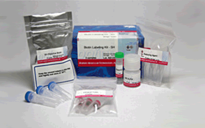
 Biotin Labeling Kit – SH
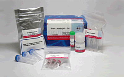
Biotin Labeling Kit – SH

抗体・タンパク質標識キット
- アミノ基でうまくいかなかった
- 感度をあげたい
-
製品コードLK10 Biotin Labeling Kit – SH
| 容 量 | メーカー希望 小売価格 |
富士フイルム 和光純薬 |
|---|---|---|
| 3 samples | ¥15,000 | 348-90941 |
| サンプル量 | 50-200 µg |
|---|---|
| 所要時間 | 3時間 |
| 標識部位 | -SH |
| 検出方法 | 顕微鏡・FCM・ウエスタンブロット・プレートリーダー |
|
・SH-Reactive Biotinと混ぜるだけで、安定な共有結合を形成する。 ・分子量50,000以上のタンパク質が標識できる。 ・付属の還元剤を用いることで遊離SH基を持たないタンパク質への標識も可能である*。 ・Filtration tubeを用いた分離操作により高い回収率で標識体が得られる。 ・付属の保存溶液でBiotin標識体の保存ができる。 |
|

| 3 samples | ・SH-Reactive Biotin ・Reducing Agent ・WS Buffer ・Reaction Buffer ・Filtration Tube |
3 tubes 3 tubes 4 ml x 1 1 ml x 1 3 tubes |
|---|
- ご購入方法
- お問い合わせ
マニュアル
-
取扱説明書
 日本語
日本語 
-
取扱説明書
 English
Biotin Labeling Kit – SHの使い方
技術情報
特長
1) 約3時間でビオチン標識体が調製できる。
2) SH-Reactive Biotinと混ぜるだけで、安定な共有結合を形成する。
3)分子量50,000以上のタンパク質が標識できる。
4)50~200 μgのタンパク質を標識可能である。
5) 付属の還元剤を用いることで、遊離SH基を持たないタンパク質への標識も可能である*。
6) Filtration Tubeを用いた分離操作により高い回収率で標識体が得られる。
7) 付属の保存溶液でBiotin標識体の保存ができる。
参考文献
1) E. Terasaka, K. Yamada, P.H. Wang, K. Hosokawa, R. Yamagiwa, K. Matsumoto, S. Ishii, T. Mori, K. Yagi, H. Sawai, H. Arai, H. Sugimoto, Y. Sugita, Y. Shiro and T. Tosha, "Dynamics of nitric oxide controlled by protein complex in bacterial system", Proc. Natl. Acad. Sci. U.S.A.., 2017, 114, (37), 9888.
2) J. Hiruma, K. Harada, A. Motoyama, Y. Okubo, T. Maeda, M. Yamamoto, M. Miyai, T. Hibino and R. Tsuboi, "Key component of inflammasome, NLRC4, was identified in the lesional epidermis of psoriatic patients", J. Dermatol.., 2018, 45, (8), 971.
3) K. Gonda, M. Watanabe, H. Tada, M. Miyashita, Y. Takahashi-Aoyama, T. Kamei, T. Ishida, S. Usami, H. Hirakawa, Y. Kakugawa, Y. Hamanaka, R. Yoshida, A. Furuta, H. Okada, H. Goda, H. Negishi, K. Takanashi, M. Takahashi, Y. Ozaki, Y. Yoshihara, Y. Nakano and N. Ohuchi, "Quantitative diagnostic imaging of cancer tissues by using phosphor-integrated dots with ultra-high brightness", Sci. Rep.., 2017, 7, (1), 7509.
4) K. Saito, M. Sakaguchi, H. Iioka, M. Matsui, H. Nakanishi, N.H. Huh and E. Kondo, "Coxsackie and adenovirus receptor is a critical regulator for the survival and growth of oral squamous carcinoma cells", Oncogene., 2014, 33, (10), 127401286.
5) K. Yamamoto, H. Murata, E.W. Putranto, K. Kataoka, A. Motoyama, T. Hibino, Y. Inoue, M. Sakaguchi and N.H. Huh, "DOCK7 is a critical regulator of the RAGE-Cdc42 signaling axis that induces formation of dendritic pseudopodia in human cancer cells", Oncol. Rep.., 2013, 29, (3), 1073.
6) M. Sakaguchi, H. Murata, K. Yamamoto, T. Ono, Y. Sakaguchi, A. Motoyama, T. Hibino, K. Kataoka and N.H. Huh, "TIRAP, an Adaptor Protein for TLR2/4, Transduces a Signal from RAGE Phosphorylated upon Ligand Binding", PLoS ONE., 2011, 6, (8), e23132.
7) M. Sakaguchi, H. Murata, Y. Aoyama, T. Hibino, E.W. Putranto, I.M. Ruma, Y. Inoue, Y. Sakaguchi, K. Yamamoto, R. Kinoshita, J. Futami, K. Kataoka, K. Iwatsuki and N.H. Huh, "DNAX-activating Protein 10 (DAP10) Membrane Adaptor Associates with Receptor for Advanced Glycation End Products (RAGE) and Modulates the RAGE-triggered Signaling Pathway in Human Keratinocytes", J. Biol. Chem.., 2014, 289, (34), 23389.
8) N. Kobayashi, K. Odaka, T. Uehara, K. Imanaka-Yoshida, Y. Kato, H. Oyama, H. Tadokoro, H. Akizawa, S. Tanada, M. Hiroe, T. Fukumura, I. Komuro, Y. Arano, T. Yoshida and T. Irie, "Toward in Vivo Imaging of Heart Disease Using a Radiolabeled Single-Chain Fv Fragment Targeting Tenascin-C", Anal. Chem.., 2011, 83, (23), 9123.
9) T. Ikeda, R. Shinohata, M. Murakami, K. Hina, S. Kamikawa, S. Hirohata, S. Kusachi, A. Tamura and S. Usui, "A rapid and precise method for measuring plasma apoE-rich HDL using polyethylene glycol and cation-exchange chromatography: a pilot study on the clinical significance of apoE-rich HDL measurements", Clin. Chim. Acta.,2017, 465, 112.
10) T. Into, M. Inomata, M. Nakashima, K. Shibata, H. Hacker and K. Matsushita, "Regulation of MyD88-Dependent Signaling Events by S Nitrosylation Retards Toll-Like Receptor Signal Transduction and Initiation of Acute-Phase Immune Responses", Mol. Cell. Biol.., 2008, 28, (4), 1338.
11) W. Nakai, T. Yoshida, D. Diez, Y. Miyatake, T. Nishibu, N. Imawaka, K. Naruse, Y. Sadamura and R. Hanayama, "A novel affinity-based method for the isolation of highly purified extracellular vesicles", Sci. Rep.., 2016, 6, 33935.
12) Y. Terasaki, T. Akuta, M. Terasaki, T. Sawa, T. Mori, T. Okamoto, M. Ozaki, M. Takeya and T. Akaike, "Guanine Nitration in Idiopathic Pulmonary Fibrosis and Its Implication for Carcinogenesis", Am. J. Respir. Crit. Care Med.., 2006, 174, (6), 665.
13) I. W. Sumardika, C. Youyi, E. Kondo, Y. Inoue, I. M. W. Ruma, H. Murata, R. Kinoshita, K. Yamamoto, S. Tomida, K. Shien, H. Sato, A. Yamauchi, J. Futami, E. W. Putranto, T. Hibino, S. Toyooka , M. Nishibori and M. Sakaguchi, "β-1,3-Galactosyl-O-Glycosyl-Glycoprotein β-1,6-N-Acetylglucosaminyltransferase 3 Increases MCAM Stability, Which Enhances S100A8/A9-Mediated Cancer Motility.", Oncol. Res., 2018, 26, (3), 431.
14) J. Iwano, D. Shinmi, K. Masuda, T. Murakami and J. Enokizono, "Impact of Different Selectivity between Soluble and Membrane-bound Forms of Carcinoembryonic Antigen (CEA) on the Target-mediated Disposition of Anti-CEA Monoclonal Antibodies.", Drug Metab. Dispos., 2019, 47, (1), 1240.
15) D. S. On, P. Chertchinnapa, Y. Shinkai, T. Kojima and H. Nakano, "Development of a dual monoclonal antibody sandwich enzyme-linked immunosorbent assay for the detection of swine influenza virus using rabbit monoclonal antibody by Ecobody technology.", J. Biosci. Bioeng., 2020, DOI:10.1016/j.jbiosc.2020.03.003.
よくある質問
-
Q
Labeling Kitで1次抗体を直接標識する利点を教えてください。
-
A
はじめて抗体標識をされる方を対象としたプロトコルを作成しております。
カスタマーサポートの視点から直接標識法の利点や実施例等を記載しておりますので、ご参照下さい。
下記リンクよりダウンロード可能です。
「はじめての抗体標識プロトコル ~カスタマーサポートの視点から~」
-
Q
サンプル溶液中の共存物は反応に影響しますか?
-
A
共存物の種類により影響することがあります。
溶液中にどのような物質が含まれるかを確認の上、状況に応じてラベル化に用いるサンプルの精製を行い、標識反応にご使用ください。<高分子:分子量1万以上>
影響する可能性があります。
高分子でSH基をもつ化合物は、Filtration Tubeでも除くことができません。 そのため標識され、蛍光性不純物として影響します。反応に使用する前に別途精製を行ってください。 一方、SH基を持たない化合物でも、高分子の不純物が多いとフィルターの目詰まりの原因になり、標識・精製操作に支障がでる可能性もあります。
*本製品に限らず他のLabeling kit に関しても同様の注意が必要です。
-
Q
低分子のタンパク質(分子量50,000以下)に標識する場合の方法を教えて下さい。
-
A
キット付属のフィルトレーションチューブは分画分子量30Kの限外濾過フィルターのため、余裕をもって50,000以上のタンパク質のご使用を推奨しております。
分子量50,000以下のタンパク質を標識される場合は、下記のような分画分子量の小さい限外濾過フィルターに変更して頂くことで、低分子のタンパク質でもラベル化可能でございます。
——————————————
PALL社 ナノセップ 3K 製品No.OD003C33
PALL社 ナノセップ 10K 製品No.OD010C33
——————————————キット同梱のフィルターを用いた場合に比べ遠心に時間を要することがございますので遠心時間はご検討下さい。
-
Q
標識後、フィルトレーションチューブを遠心してもメンブレン上に液が残る。
-
A
(1)メンブレンを目視で確認した際、メンブレンカップの淵にうっすら液が残る程度であれば、次の操作に進んで下さい。
メンブレン上に液が残っていたり、メンブレンカップを傾けて回転させたとき、液が垂れてくるようであれば、さらに遠心を8,000g で15分~30分程度行って下さい。
(2)1)の遠心操作後もメンブレン上に液が残る場合は、標識体が凝集していないか確認してください。抗体やタンパク質自身の性質によりますが、低分子標識剤を標識することで抗体やタンパク質の疎水性が増し、凝集する場合がございます。
標識体に凝集が見られる場合は、一度別のマイクロチューブに移し、遠心し上澄みをご使用ください。(抗体・タンパク質の回収量は低下いたします。)
上記でも解決しない場合は、小社カスタマーサポートまでお問い合わせください。
※フィルターの目詰まりが疑われる場合は、メンブレンフィルターを新しいものに交換すると解決する場合がございます。
代替品: PALL社製 ナノセップ 30K (メーカーコード:OD030C33)
-
Q
どのようなものが標識できますか?
-
A
分子量が「50,000以上」で[S-S]もしくは[SH]を有している化合物(抗体、蛋白質など)であれば標識できます。
量は「50~200 μg」が対象となります。
*キットに添付しているFiltration Tubeのサイズが30Kのため、低分子の化合物は精製できません。


取扱条件
| 保存条件: 冷蔵 , 取扱条件: 吸湿注意 |

関連製品
この製品に関連する研究では、下記の関連製品も使われています。
-

抗体・タンパク質標識キット
Biotin Labeling Kit – NH2
-

抗体標識キット
Ab-10 Rapid Biotin Labeling Kit

